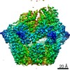
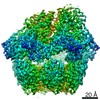
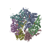
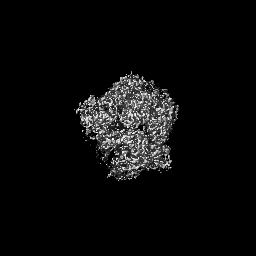
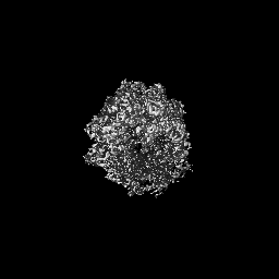
- EMDB-25502: Yeast Lon (PIM1) with endogenous substrate -
+
Open data
ID or keywords:
Loading...
-
Basic information
Entry
Database: EMDB / ID: EMD-25502
Title
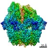
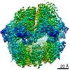
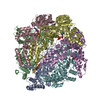
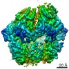
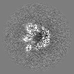
Yeast Lon (PIM1) with endogenous substrate
Map data
Sample
Complex: Cryo-EM structure of yeast Lon (PIM1) in complex with endogenous substrate
Protein or peptide: Lon protease homolog, mitochondrial
Protein or peptide: endogenous substrate
Ligand: ADENOSINE-5'-TRIPHOSPHATE
Ligand: MAGNESIUM ION
Ligand: ADENOSINE-5'-DIPHOSPHATE
Keywords
Protease / AAA+ ATPase / HYDROLASE
Function / homology
Function and homology information
regulation of mitochondrial DNA metabolic process / mitochondrial protein quality control / oxidation-dependent protein catabolic process / mitochondrial respiratory chain complex assembly / mitochondrial DNA metabolic process / endopeptidase La / Mitochondrial protein degradation / ATP-dependent peptidase activity / protein quality control for misfolded or incompletely synthesized proteins / chaperone-mediated protein complex assembly ...regulation of mitochondrial DNA metabolic process / mitochondrial protein quality control / oxidation-dependent protein catabolic process / mitochondrial respiratory chain complex assembly / mitochondrial DNA metabolic process / endopeptidase La / Mitochondrial protein degradation / ATP-dependent peptidase activity / protein quality control for misfolded or incompletely synthesized proteins / chaperone-mediated protein complex assembly / mitochondrion organization / single-stranded DNA binding / response to heat / cellular response to oxidative stress / protein folding / sequence-specific DNA binding / mitochondrial matrix / serine-type endopeptidase activity / ATP hydrolysis activity / mitochondrion / ATP binding Similarity search - Function
Lon protease homologue, chloroplastic/mitochondrial / : / Lon protease, bacterial/eukaryotic-type / Lon protease AAA+ ATPase lid domain / Peptidase S16, active site / ATP-dependent serine proteases, lon family, serine active site. / Lon proteolytic domain profile. / Peptidase S16, Lon proteolytic domain / Lon protease / Lon protease (S16) C-terminal proteolytic domain ...Lon protease homologue, chloroplastic/mitochondrial / : / Lon protease, bacterial/eukaryotic-type / Lon protease AAA+ ATPase lid domain / Peptidase S16, active site / ATP-dependent serine proteases, lon family, serine active site. / Lon proteolytic domain profile. / Peptidase S16, Lon proteolytic domain / Lon protease / Lon protease (S16) C-terminal proteolytic domain / Lon N-terminal domain profile. / Lon protease, N-terminal domain / Lon protease, N-terminal domain superfamily / ATP-dependent protease La (LON) substrate-binding domain / Found in ATP-dependent protease La (LON) / PUA-like superfamily / ATPase family associated with various cellular activities (AAA) / ATPase, AAA-type, core / Ribosomal protein S5 domain 2-type fold, subgroup / Ribosomal protein S5 domain 2-type fold / ATPases associated with a variety of cellular activities / AAA+ ATPase domain / P-loop containing nucleoside triphosphate hydrolase Similarity search - Domain/homology
National Institutes of Health/National Institute of Neurological Disorders and Stroke (NIH/NINDS)
United States
Citation
Journal: J Biol Chem / Year: 2022 Title: Cryo-EM structure of hexameric yeast Lon protease (PIM1) highlights the importance of conserved structural elements. Authors: Jie Yang / Albert S Song / R Luke Wiseman / Gabriel C Lander / Abstract: Lon protease is a conserved ATP-dependent serine protease composed of an AAA+ domain that mechanically unfolds substrates and a serine protease domain that degrades these unfolded substrates. In ...Lon protease is a conserved ATP-dependent serine protease composed of an AAA+ domain that mechanically unfolds substrates and a serine protease domain that degrades these unfolded substrates. In yeast, dysregulation of Lon protease (PIM1) attenuates lifespan and leads to gross mitochondrial morphological perturbations. Although structures of the bacterial and human Lon protease reveal a hexameric assembly, yeast PIM1 was speculated to form a heptameric assembly and is uniquely characterized by a ∼50-residue insertion between the ATPase and protease domains. To further understand the yeast-specific properties of PIM1, we determined a high-resolution cryo-electron microscopy structure of PIM1 in a substrate-translocating state. Here, we reveal that PIM1 forms a hexamer, conserved with that of bacterial and human Lon proteases, wherein the ATPase domains form a canonical closed spiral that enables pore loop residues to translocate substrates to the protease chamber. In the substrate-translocating state, PIM1 protease domains form a planar protease chamber in an active conformation and are uniquely characterized by a ∼15-residue C-terminal extension. These additional C-terminal residues form an α-helix located along the base of the protease domain. Finally, we did not observe density for the yeast-specific insertion between the ATPase and protease domains, likely due to high conformational flexibility. Biochemical studies to investigate the insertion using constructs that truncated or replaced the insertion with a glycine-serine linker suggest that the yeast-specific insertion is dispensable for PIM1's enzymatic function. Altogether, our structural and biochemical studies highlight unique components of PIM1 machinery and demonstrate evolutionary conservation of Lon protease function.
History
Deposition
Nov 24, 2021
-
Header (metadata) release
Jan 12, 2022
-
Map release
Jan 12, 2022
-
Update
May 14, 2025
-
Current status
May 14, 2025
Processing site: RCSB / Status: Released
-
Structure visualization
Movie
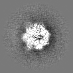
Surface view with section colored by density value
In the structure databanks used in Yorodumi, some data are registered as the other names, "COVID-19 virus" and "2019-nCoV". Here are the details of the virus and the list of structure data.
Jan 31, 2019. EMDB accession codes are about to change! (news from PDBe EMDB page)
EMDB accession codes are about to change! (news from PDBe EMDB page)
The allocation of 4 digits for EMDB accession codes will soon come to an end. Whilst these codes will remain in use, new EMDB accession codes will include an additional digit and will expand incrementally as the available range of codes is exhausted. The current 4-digit format prefixed with “EMD-” (i.e. EMD-XXXX) will advance to a 5-digit format (i.e. EMD-XXXXX), and so on. It is currently estimated that the 4-digit codes will be depleted around Spring 2019, at which point the 5-digit format will come into force.
The EM Navigator/Yorodumi systems omit the EMD- prefix.
Related info.:Q: What is EMD? / ID/Accession-code notation in Yorodumi/EM Navigator
Yorodumi is a browser for structure data from EMDB, PDB, SASBDB, etc.
This page is also the successor to EM Navigator detail page, and also detail information page/front-end page for Omokage search.
The word "yorodu" (or yorozu) is an old Japanese word meaning "ten thousand". "mi" (miru) is to see.
Related info.:EMDB / PDB / SASBDB / Comparison of 3 databanks / Yorodumi Search / Aug 31, 2016. New EM Navigator & Yorodumi / Yorodumi Papers / Jmol/JSmol / Function and homology information / Changes in new EM Navigator and Yorodumi
 Movie
Movie Controller


 Open data
 Basic information
 Map data
 Sample
 Keywords
 Function and homology information

 Authors
 United States, 1 items
  Citation
 Structure visualization
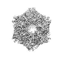
 Downloads & links
 emd_25502.png
 http://ftp.pdbj.org/pub/emdb/structures/EMD-25502
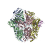


 Z (Sec.)
 Y (Row.)
 X (Col.)


 Sample components

 Processing
 Electron microscopy
 FIELD EMISSION GUN
