[English] 日本語
 Yorodumi
- EMDB-25485: In situ cryo-electron tomography reveals local cellular machineri... -
+ Open data
- Basic information
| Entry | Database: EMDB / ID: EMD-25485 | ||||||||||||||||||
|---|---|---|---|---|---|---|---|---|---|---|---|---|---|---|---|---|---|---|---|
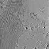
| Title | In situ cryo-electron tomography reveals local cellular machineries for axon branch development (premature branch) | ||||||||||||||||||
 Map data Map data | Tomogram of premature axon branch of hippocampal neuron. | ||||||||||||||||||
 Sample Sample |
| ||||||||||||||||||
 Keywords Keywords | In situ / cryo-ET / cryo-EM / tomography / Axon branch / Neuron / actin / microtubules / cytoskeleton / ER / ribosome / mitochondria / branching / hippocampus / CELL ADHESION | ||||||||||||||||||
| Biological species |  | ||||||||||||||||||
| Method | electron tomography / cryo EM | ||||||||||||||||||
 Authors Authors | Nedozralova H / Basnet N / Ibiricu I / Bodakuntla S / Biertumpfel C / Mizuno N | ||||||||||||||||||
| Funding support |  Germany, 5 items Germany, 5 items
| ||||||||||||||||||
 Citation Citation |  Journal: J Cell Biol / Year: 2022 Journal: J Cell Biol / Year: 2022Title: In situ cryo-electron tomography reveals local cellular machineries for axon branch development. Authors: Hana Nedozralova / Nirakar Basnet / Iosune Ibiricu / Satish Bodakuntla / Christian Biertümpfel / Naoko Mizuno /   Abstract: Neurons are highly polarized cells forming an intricate network of dendrites and axons. They are shaped by the dynamic reorganization of cytoskeleton components and cellular organelles. Axon ...Neurons are highly polarized cells forming an intricate network of dendrites and axons. They are shaped by the dynamic reorganization of cytoskeleton components and cellular organelles. Axon branching allows the formation of new paths and increases circuit complexity. However, our understanding of branch formation is sparse due to the lack of direct in-depth observations. Using in situ cellular cryo-electron tomography on primary mouse neurons, we directly visualized the remodeling of organelles and cytoskeleton structures at axon branches. Strikingly, branched areas functioned as hotspots concentrating organelles to support dynamic activities. Unaligned actin filaments assembled at the base of premature branches accompanied by filopodia-like protrusions. Microtubules and ER comigrated into preformed branches to support outgrowth together with accumulating compact, ∼500-nm mitochondria and locally clustered ribosomes. We obtained a roadmap of events supporting the hypothesis of local protein synthesis selectively taking place at axon branches, allowing them to serve as unique control hubs for axon development and downstream neural network formation. | ||||||||||||||||||
| History |
|
- Structure visualization
| Movie |
 Movie viewer Movie viewer |
|---|---|
| Supplemental images |
- Downloads & links
-EMDB archive
| Map data |  emd_25485.map.gz emd_25485.map.gz | 1.4 GB |  EMDB map data format EMDB map data format | |
|---|---|---|---|---|
| Header (meta data) |  emd-25485-v30.xml emd-25485-v30.xml emd-25485.xml emd-25485.xml | 10 KB 10 KB | Display Display |  EMDB header EMDB header |
| Images |  emd_25485.png emd_25485.png | 229.1 KB | ||
| Filedesc metadata |  emd-25485.cif.gz emd-25485.cif.gz | 4 KB | ||
| Archive directory |  http://ftp.pdbj.org/pub/emdb/structures/EMD-25485 http://ftp.pdbj.org/pub/emdb/structures/EMD-25485 ftp://ftp.pdbj.org/pub/emdb/structures/EMD-25485 ftp://ftp.pdbj.org/pub/emdb/structures/EMD-25485 | HTTPS FTP |
-Validation report
| Summary document |  emd_25485_validation.pdf.gz emd_25485_validation.pdf.gz | 562.7 KB | Display |  EMDB validaton report EMDB validaton report |
|---|---|---|---|---|
| Full document |  emd_25485_full_validation.pdf.gz emd_25485_full_validation.pdf.gz | 562.2 KB | Display | |
| Data in XML |  emd_25485_validation.xml.gz emd_25485_validation.xml.gz | 4.7 KB | Display | |
| Data in CIF |  emd_25485_validation.cif.gz emd_25485_validation.cif.gz | 5.2 KB | Display | |
| Arichive directory |  https://ftp.pdbj.org/pub/emdb/validation_reports/EMD-25485 https://ftp.pdbj.org/pub/emdb/validation_reports/EMD-25485 ftp://ftp.pdbj.org/pub/emdb/validation_reports/EMD-25485 ftp://ftp.pdbj.org/pub/emdb/validation_reports/EMD-25485 | HTTPS FTP |
-Related structure data
| Related structure data | C: citing same article ( |
|---|---|
| EM raw data |  EMPIAR-10922 (Title: Cryo-electron tomographs of mouse thalamus neurons / Data size: 71.1 EMPIAR-10922 (Title: Cryo-electron tomographs of mouse thalamus neurons / Data size: 71.1 Data #1: Reconstructed 4x binned tomograms of thalamus neurons. [reconstructed volumes])  EMPIAR-10923 (Title: Cryo-electron tomographs of mouse hippocampal neurons EMPIAR-10923 (Title: Cryo-electron tomographs of mouse hippocampal neuronsData size: 88.6 Data #1: Reconstructed 4x binned tomograms of hippocampal neurons. [reconstructed volumes]) |
- Links
Links
| EMDB pages |  EMDB (EBI/PDBe) / EMDB (EBI/PDBe) /  EMDataResource EMDataResource |
|---|
- Map
Map
| File |  Download / File: emd_25485.map.gz / Format: CCP4 / Size: 1.5 GB / Type: IMAGE STORED AS FLOATING POINT NUMBER (4 BYTES) Download / File: emd_25485.map.gz / Format: CCP4 / Size: 1.5 GB / Type: IMAGE STORED AS FLOATING POINT NUMBER (4 BYTES) | ||||||||||||||||||||||||||||||||||||||||||||||||||||||||||||
|---|---|---|---|---|---|---|---|---|---|---|---|---|---|---|---|---|---|---|---|---|---|---|---|---|---|---|---|---|---|---|---|---|---|---|---|---|---|---|---|---|---|---|---|---|---|---|---|---|---|---|---|---|---|---|---|---|---|---|---|---|---|
| Annotation | Tomogram of premature axon branch of hippocampal neuron. | ||||||||||||||||||||||||||||||||||||||||||||||||||||||||||||
| Projections & slices | Image control
Images are generated by Spider. generated in cubic-lattice coordinate | ||||||||||||||||||||||||||||||||||||||||||||||||||||||||||||
| Voxel size | X=Y=Z: 21.84 Å | ||||||||||||||||||||||||||||||||||||||||||||||||||||||||||||
| Density |
| ||||||||||||||||||||||||||||||||||||||||||||||||||||||||||||
| Symmetry | Space group: 1 | ||||||||||||||||||||||||||||||||||||||||||||||||||||||||||||
| Details | EMDB XML:
CCP4 map header:
| ||||||||||||||||||||||||||||||||||||||||||||||||||||||||||||
-Supplemental data
- Sample components
-Entire : Primary hippocampal neuron from mouse
| Entire | Name: Primary hippocampal neuron from mouse |
|---|---|
| Components |
|
-Supramolecule #1: Primary hippocampal neuron from mouse
| Supramolecule | Name: Primary hippocampal neuron from mouse / type: cell / ID: 1 / Parent: 0 / Details: Premature axon branch |
|---|---|
| Source (natural) | Organism:  |
-Experimental details
-Structure determination
| Method | cryo EM |
|---|---|
 Processing Processing | electron tomography |
| Aggregation state | cell |
- Sample preparation
| Buffer | pH: 7.5 |
|---|---|
| Grid | Model: Quantifoil R1.2/1.3 / Material: GOLD / Mesh: 300 |
| Vitrification | Cryogen name: ETHANE / Instrument: HOMEMADE PLUNGER |
| Details | embryonic neurons grown on gold EM grid |
| Sectioning | Other: NO SECTIONING |
- Electron microscopy
| Microscope | FEI TITAN KRIOS |
|---|---|
| Specialist optics | Phase plate: VOLTA PHASE PLATE / Energy filter - Name: GIF Bioquantum |
| Details | Phase plate |
| Image recording | Film or detector model: GATAN K2 SUMMIT (4k x 4k) / Average electron dose: 90.0 e/Å2 / Details: 90 e-/A^2 total dose per tilt series |
| Electron beam | Acceleration voltage: 300 kV / Electron source:  FIELD EMISSION GUN FIELD EMISSION GUN |
| Electron optics | Illumination mode: FLOOD BEAM / Imaging mode: BRIGHT FIELD / Nominal magnification: 26000 |
| Sample stage | Specimen holder model: FEI TITAN KRIOS AUTOGRID HOLDER / Cooling holder cryogen: NITROGEN |
| Experimental equipment |  Model: Titan Krios / Image courtesy: FEI Company |
- Image processing
| Final reconstruction | Algorithm: BACK PROJECTION / Software - Name:  IMOD / Number images used: 61 IMOD / Number images used: 61 |
|---|
 Movie
Movie Controller


 Z (Sec.)
 Y (Row.)
 X (Col.)


