 Movie
Movie Controller

[English] 日本語
 Yorodumi
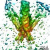
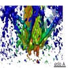
- EMDB-2545: Electron cryo-microscopy subtomogram average of a hexagon vertex ... -
+ Open data

- Basic information
| Entry | Database: EMDB / ID: EMD-2545 | |||||||||
|---|---|---|---|---|---|---|---|---|---|---|
| Title | Electron cryo-microscopy subtomogram average of a hexagon vertex in a MAC array | |||||||||
 Map data Map data | Electron cryo-microscopy subtomogram average of a hexagon vertex in a MAC array | |||||||||
 Sample Sample |
| |||||||||
 Keywords Keywords | metamorphosis / contractile / tubeworm / phage tail / extended / MAC | |||||||||
| Biological species |  Pseudoalteromonas luteoviolacea (bacteria) Pseudoalteromonas luteoviolacea (bacteria) | |||||||||
| Method | subtomogram averaging / cryo EM | |||||||||
 Authors Authors | Shikuma NJ / Pilhofer M / Weiss GL / Hadfield MG / Jensen GJ / Newman DK | |||||||||
 Citation Citation | Journal: Science / Year: 2014 Title: Marine tubeworm metamorphosis induced by arrays of bacterial phage tail-like structures. Authors: Nicholas J Shikuma / Martin Pilhofer / Gregor L Weiss / Michael G Hadfield / Grant J Jensen / Dianne K Newman /  Abstract: Many benthic marine animal populations are established and maintained by free-swimming larvae that recognize cues from surface-bound bacteria to settle and metamorphose. Larvae of the tubeworm ...Many benthic marine animal populations are established and maintained by free-swimming larvae that recognize cues from surface-bound bacteria to settle and metamorphose. Larvae of the tubeworm Hydroides elegans, an important biofouling agent, require contact with surface-bound bacteria to undergo metamorphosis; however, the mechanisms that underpin this microbially mediated developmental transition have been enigmatic. Here, we show that a marine bacterium, Pseudoalteromonas luteoviolacea, produces arrays of phage tail-like structures that trigger metamorphosis of H. elegans. These arrays comprise about 100 contractile structures with outward-facing baseplates, linked by tail fibers and a dynamic hexagonal net. Not only do these arrays suggest a novel form of bacterium-animal interaction, they provide an entry point to understanding how marine biofilms can trigger animal development. | |||||||||
| History |
|
- Structure visualization
| Movie |
 Movie viewer Movie viewer |
|---|---|
| Structure viewer | EM map: SurfViewMolmilJmol/JSmol |
| Supplemental images |

- Downloads & links
-EMDB archive
| Map data | emd_2545.map.gz | 536.9 KB | EMDB map data format | |
|---|---|---|---|---|
| Header (meta data) | emd-2545-v30.xmlemd-2545.xml | 7.7 KB 7.7 KB | Display Display | EMDB header |
| Images | PastedGraphic-8.tiff | 303.6 KB | ||
| Archive directory |  http://ftp.pdbj.org/pub/emdb/structures/EMD-2545ftp://ftp.pdbj.org/pub/emdb/structures/EMD-2545 http://ftp.pdbj.org/pub/emdb/structures/EMD-2545ftp://ftp.pdbj.org/pub/emdb/structures/EMD-2545 | HTTPS FTP |
-Related structure data
-Links
| EMDB pages | EMDB (EBI/PDBe) / EMDataResource |
|---|
-Map
| File | Download / File: emd_2545.map.gz / Format: CCP4 / Size: 635.7 KB / Type: IMAGE STORED AS FLOATING POINT NUMBER (4 BYTES) | ||||||||||||||||||||||||||||||||||||||||||||||||||||||||||||||||||||
|---|---|---|---|---|---|---|---|---|---|---|---|---|---|---|---|---|---|---|---|---|---|---|---|---|---|---|---|---|---|---|---|---|---|---|---|---|---|---|---|---|---|---|---|---|---|---|---|---|---|---|---|---|---|---|---|---|---|---|---|---|---|---|---|---|---|---|---|---|---|
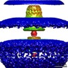
| Annotation | Electron cryo-microscopy subtomogram average of a hexagon vertex in a MAC array | ||||||||||||||||||||||||||||||||||||||||||||||||||||||||||||||||||||
| Projections & slices | Image control
Images are generated by Spider. | ||||||||||||||||||||||||||||||||||||||||||||||||||||||||||||||||||||
| Voxel size | X=Y=Z: 16.8 Å | ||||||||||||||||||||||||||||||||||||||||||||||||||||||||||||||||||||
| Density |
| ||||||||||||||||||||||||||||||||||||||||||||||||||||||||||||||||||||
| Symmetry | Space group: 1 | ||||||||||||||||||||||||||||||||||||||||||||||||||||||||||||||||||||
| Details | EMDB XML:
CCP4 map header:
| ||||||||||||||||||||||||||||||||||||||||||||||||||||||||||||||||||||
 Z (Sec.)
 Y (Row.)
 X (Col.)


-Supplemental data
- Sample components
-Entire : Culture of Pseudoalteromonas violacea cells
| Entire | Name: Culture of Pseudoalteromonas violacea cells |
|---|---|
| Components |
|
-Supramolecule #1000: Culture of Pseudoalteromonas violacea cells
| Supramolecule | Name: Culture of Pseudoalteromonas violacea cells / type: sample / ID: 1000 / Number unique components: 1 |
|---|
-Supramolecule #1: Hexagon vertex in a MAC array
| Supramolecule | Name: Hexagon vertex in a MAC array / type: organelle_or_cellular_component / ID: 1 / Oligomeric state: Structure is part of a MAC array / Recombinant expression: No / Database: NCBI |
|---|---|
| Source (natural) | Organism: Pseudoalteromonas luteoviolacea (bacteria) / Organelle: MAC array / Location in cell: cytoplasmic and extracellular |
-Experimental details
-Structure determination
| Method | cryo EM |
|---|---|
 Processing Processing | subtomogram averaging |
| Aggregation state | cell |
-Sample preparation
| Grid | Details: Quantifoil |
|---|---|
| Vitrification | Cryogen name: ETHANE-PROPANE MIXTURE / Chamber humidity: 100 % / Instrument: FEI VITROBOT MARK III |
- Electron microscopy
| Microscope | FEI POLARA 300 |
|---|---|
| Specialist optics | Energy filter - Name: GIF |
| Date | Jul 12, 2013 |
| Image recording | Category: CCD / Film or detector model: GATAN K2 (4k x 4k) |
| Electron beam | Acceleration voltage: 300 kV / Electron source:  FIELD EMISSION GUN FIELD EMISSION GUN |
| Electron optics | Illumination mode: FLOOD BEAM / Imaging mode: BRIGHT FIELD |
| Sample stage | Specimen holder model: OTHER |
| Experimental equipment |  Model: Tecnai Polara / Image courtesy: FEI Company |
-Image processing
| Details | PEET |
|---|---|
| Final reconstruction | Algorithm: OTHER / Software - Name: ETOMO / Number subtomograms used: 60 |
