 ムービー
ムービー コントローラー


+ データを開く

- 基本情報
基本情報
| 登録情報 | データベース: EMDB / ID: EMD-2521 | |||||||||
|---|---|---|---|---|---|---|---|---|---|---|
| タイトル | Flagellar hook basal body in the tomogram of Salmonella mini-cell | |||||||||
 マップデータ マップデータ | Flagellar hook basal body from Salmonella mini-cell | |||||||||
 試料 試料 |
| |||||||||
 キーワード キーワード | Flagellar motor / Type III secretion system / Salmonella typhimurium | |||||||||
| 生物種 |  Salmonella enterica subsp. enterica serovar Typhimurium (サルモネラ菌) Salmonella enterica subsp. enterica serovar Typhimurium (サルモネラ菌) | |||||||||
| 手法 | サブトモグラム平均法 / クライオ電子顕微鏡法 / 解像度: 45.0 Å | |||||||||
 データ登録者 データ登録者 | Kawamoto A / Morimoto VY / Miyata T / Minamino T / Hughes TK / Kato T / Namba K | |||||||||
 引用 引用 | ジャーナル: Sci Rep / 年: 2013 タイトル: Common and distinct structural features of Salmonella injectisome and flagellar basal body. 著者: Akihiro Kawamoto / Yusuke V Morimoto / Tomoko Miyata / Tohru Minamino / Kelly T Hughes / Takayuki Kato / Keiichi Namba /  要旨: Bacterial pathogens use an injectisome to deliver virulence proteins into eukaryotic host cells. The bacterial flagellum and injectisome export their component proteins for self-assembly. These two ...Bacterial pathogens use an injectisome to deliver virulence proteins into eukaryotic host cells. The bacterial flagellum and injectisome export their component proteins for self-assembly. These two systems show high structural similarities and are classified as the type III secretion system, but it remains elusive how similar they are in situ because the structures of these complexes isolated from cells and visualized by electron cryomicroscopy have shown only the export channel and housing for the export apparatus. Here we report in situ structures of Salmonella injectisome and flagellum by electron cryotomography. The injectisome lacks the flagellar basal body C-ring, but a wing-like disc and a globular density corresponding to the export gate platform and ATPase hexamer ring, respectively, are stably attached through thin connectors, revealing yet unidentified common architectures of the two systems. The ATPase ring is far from the disc, suggesting that both apparatuses are observed in an export-off state. | |||||||||
| 履歴 |
|
- 構造の表示
構造の表示
| ムービー |
 ムービービューア ムービービューア |
|---|---|
| 構造ビューア | EMマップ: SurfViewMolmilJmol/JSmol |
| 添付画像 |

- ダウンロードとリンク
-EMDBアーカイブ
| マップデータ | emd_2521.map.gz | 1.7 MB | EMDBマップデータ形式 | |
|---|---|---|---|---|
| ヘッダ (付随情報) | emd-2521-v30.xmlemd-2521.xml | 8.1 KB 8.1 KB | 表示 表示 | EMDBヘッダ |
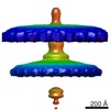
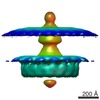
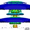
| 画像 | 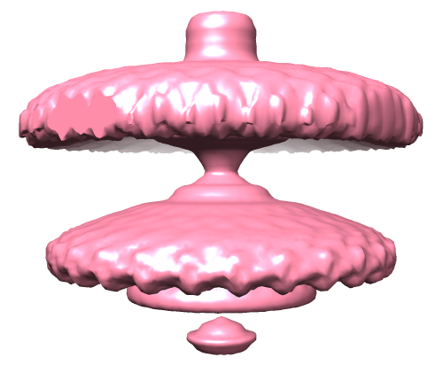 EMD-2521-1.png EMD-2521-1.png | 103.6 KB | ||
| アーカイブディレクトリ |  http://ftp.pdbj.org/pub/emdb/structures/EMD-2521ftp://ftp.pdbj.org/pub/emdb/structures/EMD-2521 http://ftp.pdbj.org/pub/emdb/structures/EMD-2521ftp://ftp.pdbj.org/pub/emdb/structures/EMD-2521 | HTTPS FTP |
-検証レポート
| 文書・要旨 | emd_2521_validation.pdf.gz | 215.4 KB | 表示 | EMDB検証レポート |
|---|---|---|---|---|
| 文書・詳細版 | emd_2521_full_validation.pdf.gz | 214.6 KB | 表示 | |
| XML形式データ | emd_2521_validation.xml.gz | 5.5 KB | 表示 | |
| アーカイブディレクトリ | https://ftp.pdbj.org/pub/emdb/validation_reports/EMD-2521ftp://ftp.pdbj.org/pub/emdb/validation_reports/EMD-2521 | HTTPS FTP |
-関連構造データ
-リンク
| EMDBのページ | EMDB (EBI/PDBe) / EMDataResource |
|---|
-マップ
| ファイル | ダウンロード / ファイル: emd_2521.map.gz / 形式: CCP4 / 大きさ: 2.1 MB / タイプ: IMAGE STORED AS FLOATING POINT NUMBER (4 BYTES) | ||||||||||||||||||||||||||||||||||||||||||||||||||||||||||||||||||||
|---|---|---|---|---|---|---|---|---|---|---|---|---|---|---|---|---|---|---|---|---|---|---|---|---|---|---|---|---|---|---|---|---|---|---|---|---|---|---|---|---|---|---|---|---|---|---|---|---|---|---|---|---|---|---|---|---|---|---|---|---|---|---|---|---|---|---|---|---|---|
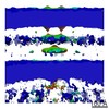
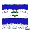
| 注釈 | Flagellar hook basal body from Salmonella mini-cell | ||||||||||||||||||||||||||||||||||||||||||||||||||||||||||||||||||||
| 投影像・断面図 | 画像のコントロール
画像は Spider により作成 | ||||||||||||||||||||||||||||||||||||||||||||||||||||||||||||||||||||
| ボクセルのサイズ | X=Y=Z: 11.4 Å | ||||||||||||||||||||||||||||||||||||||||||||||||||||||||||||||||||||
| 密度 |
| ||||||||||||||||||||||||||||||||||||||||||||||||||||||||||||||||||||
| 対称性 | 空間群: 1 | ||||||||||||||||||||||||||||||||||||||||||||||||||||||||||||||||||||
| 詳細 | EMDB XML:
CCP4マップ ヘッダ情報:
| ||||||||||||||||||||||||||||||||||||||||||||||||||||||||||||||||||||
 Z (Sec.)
 Y (Row.)
 X (Col.)


-添付データ
- 試料の構成要素
-全体 : Flagellar hook basal body from Salmonella typhimurium mini-cell i...
| 全体 | 名称: Flagellar hook basal body from Salmonella typhimurium mini-cell in situ |
|---|---|
| 要素 |
|
-超分子 #1000: Flagellar hook basal body from Salmonella typhimurium mini-cell i...
| 超分子 | 名称: Flagellar hook basal body from Salmonella typhimurium mini-cell in situ タイプ: sample / ID: 1000 / Number unique components: 1 |
|---|
-超分子 #1: flagellar hook basal body
| 超分子 | 名称: flagellar hook basal body / タイプ: organelle_or_cellular_component / ID: 1 / Name.synonym: flagellar motor / 組換発現: No |
|---|---|
| 由来(天然) | 生物種: Salmonella enterica subsp. enterica serovar Typhimurium (サルモネラ菌) 細胞中の位置: Plasma membrane |
-実験情報
-構造解析
| 手法 | クライオ電子顕微鏡法 |
|---|---|
 解析 解析 | サブトモグラム平均法 |
| 試料の集合状態 | cell |
-試料調製
| 緩衝液 | 詳細: M9 medium (17.1g Na2HPO4-12H2O, 3g KH2PO4, 0.5g NaCl, 1g NH4Cl, 0.2%glycerol, 1% tryptone, 1mM MgSO4 per litre) |
|---|---|
| グリッド | 詳細: Quantifoil molybdenum 200 mesh R0.6/1.0 grid with thin carbon support |
| 凍結 | 凍結剤: ETHANE / チャンバー内湿度: 100 % / 装置: FEI VITROBOT MARK II |
- 電子顕微鏡法
| 顕微鏡 | FEI TITAN KRIOS |
|---|---|
| 温度 | 最低: 80 K |
| 日付 | 2012年12月6日 |
| 撮影 | カテゴリ: CCD フィルム・検出器のモデル: FEI FALCON I (4k x 4k) 平均電子線量: 200 e/Å2 |
| 電子線 | 加速電圧: 300 kV / 電子線源:  FIELD EMISSION GUN FIELD EMISSION GUN |
| 電子光学系 | 倍率(補正後): 49030 / 照射モード: FLOOD BEAM / 撮影モード: BRIGHT FIELD / Cs: 2.7 mm / 最大 デフォーカス(公称値): 7.0 µm / 最小 デフォーカス(公称値): 4.0 µm / 倍率(公称値): 29000 |
| 試料ステージ | 試料ホルダーモデル: FEI TITAN KRIOS AUTOGRID HOLDER Tilt series - Axis1 - Min angle: -70 ° / Tilt series - Axis1 - Max angle: 70 ° |
| 実験機器 |  モデル: Titan Krios / 画像提供: FEI Company |
-画像解析
| 最終 再構成 | 想定した対称性 - 点群: C26 (26回回転対称) / アルゴリズム: OTHER / 解像度のタイプ: BY AUTHOR / 解像度: 45.0 Å / 解像度の算出法: OTHER / ソフトウェア - 名称: EMAN / 使用したサブトモグラム数: 48 |
|---|
