 Movie
Movie Controller

[English] 日本語
 Yorodumi
- EMDB-25096: Electron cryo-tomography of CHIKV assembly intermediates in human... -
+ Open data

- Basic information
| Entry |  | |||||||||
|---|---|---|---|---|---|---|---|---|---|---|
| Title | Electron cryo-tomography of CHIKV assembly intermediates in human U2OS cells | |||||||||
 Map data Map data | CHIKV assembly/budding intermediate from virus-infected human U2OS cells | |||||||||
 Sample Sample |
| |||||||||
 Keywords Keywords | alphavirus / enveloped virus / virus assembly and budding / cryogenic electron tomography / subtomogram averaging / VIRUS | |||||||||
| Biological species |   Chikungunya virus Chikungunya virus | |||||||||
| Method | subtomogram averaging / cryo EM / Resolution: 23.0 Å | |||||||||
 Authors Authors | Chmielewski D / Schmid MF / Simmons G / Jin J / Chiu W | |||||||||
| Funding support |  United States, 1 items United States, 1 items
| |||||||||
 Citation Citation | Journal: To Be Published Title: In situ Alphavirus Assembly and Budding Mechanism Revealed by Cellular CryoET Authors: Chmielewski D / Schmid M / Simmons G / Jin J / Chiu W | |||||||||
| History |
|
- Structure visualization
| Supplemental images |
|---|

- Downloads & links
-EMDB archive
| Map data | emd_25096.map.gz | 43.8 MB |  EMDB map data format EMDB map data format | |
|---|---|---|---|---|
| Header (meta data) | emd-25096-v30.xmlemd-25096.xml | 8.7 KB 8.7 KB | Display Display | EMDB header |
| Images | 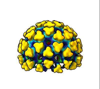 emd_25096.png emd_25096.png | 83.4 KB | ||
| Filedesc metadata | emd-25096.cif.gz | 3.7 KB | ||
| Archive directory |  http://ftp.pdbj.org/pub/emdb/structures/EMD-25096ftp://ftp.pdbj.org/pub/emdb/structures/EMD-25096 http://ftp.pdbj.org/pub/emdb/structures/EMD-25096ftp://ftp.pdbj.org/pub/emdb/structures/EMD-25096 | HTTPS FTP |
-Validation report
| Summary document | emd_25096_validation.pdf.gz | 391.5 KB | Display | EMDB validaton report |
|---|---|---|---|---|
| Full document | emd_25096_full_validation.pdf.gz | 391.1 KB | Display | |
| Data in XML | emd_25096_validation.xml.gz | 7.2 KB | Display | |
| Data in CIF | emd_25096_validation.cif.gz | 8.2 KB | Display | |
| Arichive directory | https://ftp.pdbj.org/pub/emdb/validation_reports/EMD-25096ftp://ftp.pdbj.org/pub/emdb/validation_reports/EMD-25096 | HTTPS FTP |
-Related structure data
| Related structure data | C: citing same article ( |
|---|


-Links
| EMDB pages | EMDB (EBI/PDBe) / EMDataResource |
|---|
-Map
| File | Download / File: emd_25096.map.gz / Format: CCP4 / Size: 125 MB / Type: IMAGE STORED AS FLOATING POINT NUMBER (4 BYTES) | ||||||||||||||||||||
|---|---|---|---|---|---|---|---|---|---|---|---|---|---|---|---|---|---|---|---|---|---|
| Annotation | CHIKV assembly/budding intermediate from virus-infected human U2OS cells | ||||||||||||||||||||
| Voxel size | X=Y=Z: 3.54 Å | ||||||||||||||||||||
| Density |
| ||||||||||||||||||||
| Symmetry | Space group: 1 | ||||||||||||||||||||
| Details | EMDB XML:
|
-Supplemental data
- Sample components
-Entire : Chikungunya virus
| Entire | Name: Chikungunya virus |
|---|---|
| Components |
|
-Supramolecule #1: Chikungunya virus
| Supramolecule | Name: Chikungunya virus / type: virus / ID: 1 / Parent: 0 Details: CHIKV assembly/budding intermediate from virus-infected human U2OS cells NCBI-ID: 37124 / Sci species name: Chikungunya virus / Sci species strain: 181/clone 25 / Virus type: VIRION / Virus isolate: STRAIN / Virus enveloped: Yes / Virus empty: No |
|---|---|
| Molecular weight | Theoretical: 65 MDa |
| Virus shell | Shell ID: 1 / Diameter: 700.0 Å / T number (triangulation number): 4 |
-Experimental details
-Structure determination
| Method | cryo EM |
|---|---|
 Processing Processing | subtomogram averaging |
| Aggregation state | particle |
-Sample preparation
| Buffer | pH: 8 |
|---|---|
| Vitrification | Cryogen name: ETHANE |
- Electron microscopy
| Microscope | TFS TALOS |
|---|---|
| Image recording | Film or detector model: GATAN K2 SUMMIT (4k x 4k) / Detector mode: COUNTING / Average electron dose: 1.8 e/Å2 |
| Electron beam | Acceleration voltage: 200 kV / Electron source:  FIELD EMISSION GUN FIELD EMISSION GUN |
| Electron optics | Illumination mode: FLOOD BEAM / Imaging mode: BRIGHT FIELD |
-Image processing
| Final reconstruction | Applied symmetry - Point group: C5 (5 fold cyclic) / Resolution.type: BY AUTHOR / Resolution: 23.0 Å / Resolution method: FSC 0.143 CUT-OFF / Number subtomograms used: 75 |
|---|---|
| Extraction | Number tomograms: 185 / Number images used: 75 |
| Final angle assignment | Type: PROJECTION MATCHING |
