 Movie
Movie Controller

[English] 日本語
 Yorodumi
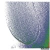
- EMDB-25047: Tomogram of mouse stereocilia containing PCDH15 molecules labeled... -
+ Open data

- Basic information
| Entry | Database: EMDB / ID: EMD-25047 | |||||||||
|---|---|---|---|---|---|---|---|---|---|---|
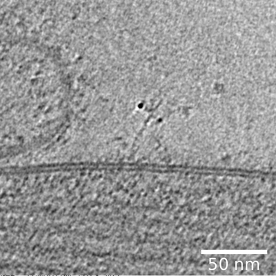
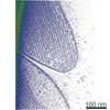
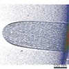
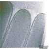
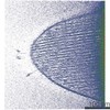
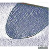
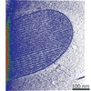
| Title | Tomogram of mouse stereocilia containing PCDH15 molecules labeled with 39G7-AuNPs | |||||||||
 Map data Map data | Tomogram of mouse stereocilia containing PCDH15 molecules labeled with 39G7-AuNPs | |||||||||
 Sample Sample |
| |||||||||
| Biological species |  | |||||||||
| Method | electron tomography / cryo EM | |||||||||
 Authors Authors | Elferich J / Clark S / Ge J / Goehring A / Matsui A / Gouaux E | |||||||||
| Funding support |  United States, 1 items United States, 1 items
| |||||||||
 Citation Citation | Journal: Elife / Year: 2021 Title: Molecular structures and conformations of protocadherin-15 and its complexes on stereocilia elucidated by cryo-electron tomography. Authors: Johannes Elferich / Sarah Clark / Jingpeng Ge / April Goehring / Aya Matsui / Eric Gouaux / Abstract: Mechanosensory transduction (MT), the conversion of mechanical stimuli into electrical signals, underpins hearing and balance and is carried out within hair cells in the inner ear. Hair cells harbor ...Mechanosensory transduction (MT), the conversion of mechanical stimuli into electrical signals, underpins hearing and balance and is carried out within hair cells in the inner ear. Hair cells harbor actin-filled stereocilia, arranged in rows of descending heights, where the tips of stereocilia are connected to their taller neighbors by a filament composed of protocadherin 15 (PCDH15) and cadherin 23 (CDH23), deemed the 'tip link.' Tension exerted on the tip link opens an ion channel at the tip of the shorter stereocilia, thus converting mechanical force into an electrical signal. While biochemical and structural studies have provided insights into the molecular composition and structure of isolated portions of the tip link, the architecture, location, and conformational states of intact tip links, on stereocilia, remains unknown. Here, we report in situ cryo-electron microscopy imaging of the tip link in mouse stereocilia. We observe individual PCDH15 molecules at the tip and shaft of stereocilia and determine their stoichiometry, conformational heterogeneity, and their complexes with other filamentous proteins, perhaps including CDH23. The PCDH15 complexes occur in clusters, frequently with more than one copy of PCDH15 at the tip of stereocilia, suggesting that tip links might consist of more than one copy of PCDH15 complexes and, by extension, might include multiple MT complexes. #1: Journal: Biorxiv / Year: 2021Title: Molecular structure and conformation of stereocilia tip-links elucidated by cryo-electron tomography Authors: Elferich J / Clark S / Ge J / Goehring A / Matsui A / Gouaux E | |||||||||
| History |
|
- Structure visualization
| Movie |
 Movie viewer Movie viewer |
|---|---|
| Supplemental images |
- Downloads & links
-EMDB archive
| Map data | emd_25047.map.gz | 633.6 MB | EMDB map data format | |
|---|---|---|---|---|
| Header (meta data) | emd-25047-v30.xmlemd-25047.xml | 13.5 KB 13.5 KB | Display Display | EMDB header |
| Images |  emd_25047.png emd_25047.png | 140.7 KB | ||
| Archive directory |  http://ftp.pdbj.org/pub/emdb/structures/EMD-25047ftp://ftp.pdbj.org/pub/emdb/structures/EMD-25047 http://ftp.pdbj.org/pub/emdb/structures/EMD-25047ftp://ftp.pdbj.org/pub/emdb/structures/EMD-25047 | HTTPS FTP |
-Related structure data
| Related structure data | C: citing same article ( |
|---|---|
| EM raw data | EMPIAR-10820 (Title: Molecular structure and conformation of stereocilia tip-links elucidated by cryo-electron tomography Data size: 58.5 Data #1: Tiltseries of mPCDH15/39G7-AuNP complex [tilt series] Data #2: Tiltseries of mouse stereocilia containing PCDH15 molecules labeled with 39G7-AuNPs [tilt series] Data #3: Tiltseries of mouse stereocilia containing PCDH15 molecules labeled with 39G7-AuNPs [tilt series] Data #4: Tiltseries of mouse stereocilia containing PCDH15 molecules labeled with 39G7-AuNPs [tilt series] Data #5: Tiltseries of mouse stereocilia containing PCDH15 molecules labeled with 39G7-AuNPs [tilt series] Data #6: Tiltseries of mouse stereocilia containing PCDH15 molecules labeled with 39G7-AuNPs [tilt series] Data #7: Tiltseries of mouse stereocilia containing PCDH15 molecules labeled with 39G7-AuNPs [tilt series] Data #8: Tiltseries of mouse stereocilia containing PCDH15 molecules labeled with 39G7-AuNPs [tilt series] Data #9: Tiltseries of mouse stereocilia containing PCDH15 molecules labeled with 39G7-AuNPs [tilt series] Data #10: Tiltseries of mouse stereocilia containing PCDH15 molecules labeled with 39G7-AuNPs [tilt series] Data #11: Tiltseries of mouse stereocilia containing PCDH15 molecules labeled with 39G7-AuNPs [tilt series] Data #12: Tiltseries of mouse stereocilia containing PCDH15 molecules labeled with 39G7-AuNPs [tilt series] Data #13: Tiltseries of mouse stereocilia containing PCDH15 molecules labeled with 39G7-AuNPs [tilt series] Data #14: Tiltseries of mouse stereocilia containing PCDH15 molecules labeled with 39G7-AuNPs [tilt series] Data #15: Tiltseries of mouse stereocilia containing PCDH15 molecules labeled with 39G7-AuNPs [tilt series] Data #16: Tiltseries of mouse stereocilia containing PCDH15 molecules labeled with 39G7-AuNPs [tilt series]) EMPIAR-10898 (Title: Cryo-electron tilt series of mouse stereocilia / Data size: 1.9 TBData #1: Unaligned tilt series of mouse stereocilia [tilt series]) |

-Links
| EMDB pages | EMDB (EBI/PDBe) / EMDataResource |
|---|
-Map
| File | Download / File: emd_25047.map.gz / Format: CCP4 / Size: 746.9 MB / Type: IMAGE STORED AS SIGNED BYTE | ||||||||||||||||||||||||||||||||||||||||||||||||||||||||||||||||||||
|---|---|---|---|---|---|---|---|---|---|---|---|---|---|---|---|---|---|---|---|---|---|---|---|---|---|---|---|---|---|---|---|---|---|---|---|---|---|---|---|---|---|---|---|---|---|---|---|---|---|---|---|---|---|---|---|---|---|---|---|---|---|---|---|---|---|---|---|---|---|
| Annotation | Tomogram of mouse stereocilia containing PCDH15 molecules labeled with 39G7-AuNPs | ||||||||||||||||||||||||||||||||||||||||||||||||||||||||||||||||||||
| Voxel size | X=Y=Z: 6.611 Å | ||||||||||||||||||||||||||||||||||||||||||||||||||||||||||||||||||||
| Density |
| ||||||||||||||||||||||||||||||||||||||||||||||||||||||||||||||||||||
| Symmetry | Space group: 1 | ||||||||||||||||||||||||||||||||||||||||||||||||||||||||||||||||||||
| Details | EMDB XML:
CCP4 map header:
| ||||||||||||||||||||||||||||||||||||||||||||||||||||||||||||||||||||
-Supplemental data
- Sample components
-Entire : Stereocilia labeled with 39G7-AuNP
| Entire | Name: Stereocilia labeled with 39G7-AuNP |
|---|---|
| Components |
|
-Supramolecule #1: Stereocilia labeled with 39G7-AuNP
| Supramolecule | Name: Stereocilia labeled with 39G7-AuNP / type: organelle_or_cellular_component / ID: 1 / Parent: 0 |
|---|
-Supramolecule #2: Mouse stereocilia
| Supramolecule | Name: Mouse stereocilia / type: organelle_or_cellular_component / ID: 2 / Parent: 1 |
|---|---|
| Source (natural) | Organism: |
-Supramolecule #3: 39G7-AuNP conjugate
| Supramolecule | Name: 39G7-AuNP conjugate / type: complex / ID: 3 / Parent: 1 |
|---|---|
| Source (natural) | Organism: |
| Recombinant expression | Organism:   Spodoptera frugiperda (fall armyworm) Spodoptera frugiperda (fall armyworm) |
-Experimental details
-Structure determination
| Method | cryo EM |
|---|---|
 Processing Processing | electron tomography |
| Aggregation state | tissue |
-Sample preparation
| Buffer | pH: 7.4 / Details: DMEM/F12 |
|---|---|
| Grid | Model: C-flat-2/2 / Material: COPPER / Mesh: 200 / Support film - Material: CARBON / Support film - topology: HOLEY / Pretreatment - Type: GLOW DISCHARGE / Details: Coated with Polylysine |
| Vitrification | Cryogen name: ETHANE-PROPANE / Instrument: HOMEMADE PLUNGER |
| Sectioning | Other: NO SECTIONING |
| Fiducial marker | Manufacturer: PELCO / Diameter: 10 nm |
- Electron microscopy
| Microscope | FEI TITAN KRIOS |
|---|---|
| Image recording | Film or detector model: GATAN K3 BIOQUANTUM (6k x 4k) / Number real images: 41 / Average electron dose: 3.0 e/Å2 |
| Electron beam | Acceleration voltage: 300 kV / Electron source:  FIELD EMISSION GUN FIELD EMISSION GUN |
| Electron optics | Illumination mode: FLOOD BEAM / Imaging mode: BRIGHT FIELD / Cs: 2.7 mm |
| Experimental equipment |  Model: Titan Krios / Image courtesy: FEI Company |
-Image processing
| Final reconstruction | Algorithm: BACK PROJECTION / Software - Name: TomoAlign (ver. Jan2019) / Details: Filtered with SIRT-like filter / Number images used: 41 |
|---|---|
| CTF correction | Software - Name: IMOD |
