+ Open data
- Basic information
| Entry |  | |||||||||
|---|---|---|---|---|---|---|---|---|---|---|
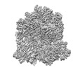
| Title | Damaged 70S ribosome with PrfH bound | |||||||||
 Map data Map data | ||||||||||
 Sample Sample |
| |||||||||
 Keywords Keywords | PrfH / damaged ribosome / ribotoxin / RIBOSOME | |||||||||
| Function / homology |  Function and homology information Function and homology informationtranslation release factor activity / positive regulation of ribosome biogenesis / DnaA-L2 complex / negative regulation of DNA-templated DNA replication initiation / assembly of large subunit precursor of preribosome / cytosolic ribosome assembly / regulation of cell growth / mRNA 5'-UTR binding / ribosome biogenesis / large ribosomal subunit ...translation release factor activity / positive regulation of ribosome biogenesis / DnaA-L2 complex / negative regulation of DNA-templated DNA replication initiation / assembly of large subunit precursor of preribosome / cytosolic ribosome assembly / regulation of cell growth / mRNA 5'-UTR binding / ribosome biogenesis / large ribosomal subunit / transferase activity / ribosome binding / ribosomal small subunit assembly / small ribosomal subunit / 5S rRNA binding / ribosomal large subunit assembly / cytosolic small ribosomal subunit / large ribosomal subunit rRNA binding / small ribosomal subunit rRNA binding / cytosolic large ribosomal subunit / cytoplasmic translation / tRNA binding / negative regulation of translation / rRNA binding / ribosome / structural constituent of ribosome / translation / ribonucleoprotein complex / mRNA binding / RNA binding / zinc ion binding / metal ion binding / membrane / cytosol / cytoplasm Similarity search - Function | |||||||||
| Biological species |  | |||||||||
| Method | single particle reconstruction / cryo EM / Resolution: 2.55 Å | |||||||||
 Authors Authors | Tian Y / Zeng F | |||||||||
| Funding support |  United States, 1 items United States, 1 items
| |||||||||
 Citation Citation |  Journal: Proc Natl Acad Sci U S A / Year: 2022 Journal: Proc Natl Acad Sci U S A / Year: 2022Title: Sequential rescue and repair of stalled and damaged ribosome by bacterial PrfH and RtcB. Authors: Yannan Tian / Fuxing Zeng / Adrika Raybarman / Shirin Fatma / Amy Carruthers / Qingrong Li / Raven H Huang /   Abstract: RtcB is involved in transfer RNA (tRNA) splicing in archaeal and eukaryotic organisms. However, most RtcBs are found in bacteria, whose tRNAs have no introns. Because tRNAs are the substrates of ...RtcB is involved in transfer RNA (tRNA) splicing in archaeal and eukaryotic organisms. However, most RtcBs are found in bacteria, whose tRNAs have no introns. Because tRNAs are the substrates of archaeal and eukaryotic RtcB, it is assumed that bacterial RtcBs are for repair of damaged tRNAs. Here, we show that a subset of bacterial RtcB, denoted RtcB2 herein, specifically repair ribosomal damage in the decoding center. To access the damage site for repair, however, the damaged 70S ribosome needs to be dismantled first, and this is accomplished by bacterial PrfH. Peptide-release assays revealed that PrfH is only active with the damaged 70S ribosome but not with the intact one. A 2.55-Å cryo-electron microscopy structure of PrfH in complex with the damaged 70S ribosome provides molecular insight into PrfH discriminating between the damaged and the intact ribosomes via specific recognition of the cleaved 3'-terminal nucleotide. RNA repair assays demonstrated that RtcB2 efficiently repairs the damaged 30S ribosomal subunit but not the damaged tRNAs. Cell-based assays showed that the RtcB2-PrfH pair reverse the damage inflicted by ribosome-specific ribotoxins in vivo. Thus, our combined biochemical, structural, and cell-based studies have uncovered a bacterial defense system specifically evolved to reverse the lethal ribosomal damage in the decoding center for cell survival. | |||||||||
| History |
|
- Structure visualization
| Supplemental images |
|---|
- Downloads & links
-EMDB archive
| Map data |  emd_24944.map.gz emd_24944.map.gz | 202.5 MB |  EMDB map data format EMDB map data format | |
|---|---|---|---|---|
| Header (meta data) |  emd-24944-v30.xml emd-24944-v30.xml emd-24944.xml emd-24944.xml | 79.9 KB 79.9 KB | Display Display |  EMDB header EMDB header |
| FSC (resolution estimation) |  emd_24944_fsc.xml emd_24944_fsc.xml | 13.5 KB | Display |  FSC data file FSC data file |
| Images |  emd_24944.png emd_24944.png | 134.5 KB | ||
| Filedesc metadata |  emd-24944.cif.gz emd-24944.cif.gz | 15.5 KB | ||
| Archive directory |  http://ftp.pdbj.org/pub/emdb/structures/EMD-24944 http://ftp.pdbj.org/pub/emdb/structures/EMD-24944 ftp://ftp.pdbj.org/pub/emdb/structures/EMD-24944 ftp://ftp.pdbj.org/pub/emdb/structures/EMD-24944 | HTTPS FTP |
-Related structure data
| Related structure data |  7sa4MC M: atomic model generated by this map C: citing same article ( |
|---|---|
| Similar structure data | Similarity search - Function & homology  F&H Search F&H Search |
- Links
Links
| EMDB pages |  EMDB (EBI/PDBe) / EMDB (EBI/PDBe) /  EMDataResource EMDataResource |
|---|---|
| Related items in Molecule of the Month |
- Map
Map
| File |  Download / File: emd_24944.map.gz / Format: CCP4 / Size: 216 MB / Type: IMAGE STORED AS FLOATING POINT NUMBER (4 BYTES) Download / File: emd_24944.map.gz / Format: CCP4 / Size: 216 MB / Type: IMAGE STORED AS FLOATING POINT NUMBER (4 BYTES) | ||||||||||||||||||||||||||||||||||||
|---|---|---|---|---|---|---|---|---|---|---|---|---|---|---|---|---|---|---|---|---|---|---|---|---|---|---|---|---|---|---|---|---|---|---|---|---|---|
| Projections & slices | Image control
Images are generated by Spider. | ||||||||||||||||||||||||||||||||||||
| Voxel size | X=Y=Z: 1.05 Å | ||||||||||||||||||||||||||||||||||||
| Density |
| ||||||||||||||||||||||||||||||||||||
| Symmetry | Space group: 1 | ||||||||||||||||||||||||||||||||||||
| Details | EMDB XML:
|
-Supplemental data
- Sample components
+Entire : Damaged 70S with PrfH
+Supramolecule #1: Damaged 70S with PrfH
+Supramolecule #2: 50S
+Supramolecule #3: 30S
+Supramolecule #4: tRNA
+Supramolecule #5: Putative peptide chain release factor homolog
+Macromolecule #1: 23S ribosomal RNA
+Macromolecule #2: 16S ribosomal RNA
+Macromolecule #3: 5S ribosomal RNA
+Macromolecule #4: P-tRNA, E-tRNA
+Macromolecule #5: mRNA
+Macromolecule #6: Peptide chain release factor H
+Macromolecule #7: 50S ribosomal protein L2
+Macromolecule #8: 50S ribosomal protein L3
+Macromolecule #9: 50S ribosomal protein L4
+Macromolecule #10: 50S ribosomal protein L5
+Macromolecule #11: 50S ribosomal protein L6
+Macromolecule #12: 50S ribosomal protein L9
+Macromolecule #13: 50S ribosomal protein L10
+Macromolecule #14: 50S ribosomal protein L11
+Macromolecule #15: 50S ribosomal protein L13
+Macromolecule #16: 50S ribosomal protein L14
+Macromolecule #17: 50S ribosomal protein L15
+Macromolecule #18: 50S ribosomal protein L16
+Macromolecule #19: 50S ribosomal protein L17
+Macromolecule #20: 50S ribosomal protein L18
+Macromolecule #21: 50S ribosomal protein L19
+Macromolecule #22: 50S ribosomal protein L20
+Macromolecule #23: 50S ribosomal protein L21
+Macromolecule #24: 50S ribosomal protein L22
+Macromolecule #25: 50S ribosomal protein L23
+Macromolecule #26: 50S ribosomal protein L24
+Macromolecule #27: 50S ribosomal protein L25
+Macromolecule #28: 50S ribosomal protein L27
+Macromolecule #29: 50S ribosomal protein L28
+Macromolecule #30: 50S ribosomal protein L29
+Macromolecule #31: 50S ribosomal protein L30
+Macromolecule #32: 50S ribosomal protein L31
+Macromolecule #33: 50S ribosomal protein L32
+Macromolecule #34: 50S ribosomal protein L33
+Macromolecule #35: 50S ribosomal protein L34
+Macromolecule #36: 50S ribosomal protein L35
+Macromolecule #37: 50S ribosomal protein L36
+Macromolecule #38: 30S ribosomal protein S2
+Macromolecule #39: 30S ribosomal protein S3
+Macromolecule #40: 30S ribosomal protein S4
+Macromolecule #41: 30S ribosomal protein S5
+Macromolecule #42: 30S ribosomal protein S6
+Macromolecule #43: 30S ribosomal protein S7
+Macromolecule #44: 30S ribosomal protein S8
+Macromolecule #45: 30S ribosomal protein S9
+Macromolecule #46: 30S ribosomal protein S10
+Macromolecule #47: 30S ribosomal protein S11
+Macromolecule #48: 30S ribosomal protein S12
+Macromolecule #49: 30S ribosomal protein S13
+Macromolecule #50: 30S ribosomal protein S14
+Macromolecule #51: 30S ribosomal protein S15
+Macromolecule #52: 30S ribosomal protein S16
+Macromolecule #53: 30S ribosomal protein S17
+Macromolecule #54: 30S ribosomal protein S18
+Macromolecule #55: 30S ribosomal protein S19
+Macromolecule #56: 30S ribosomal protein S20
+Macromolecule #57: 30S ribosomal protein S21
+Macromolecule #58: MAGNESIUM ION
+Macromolecule #59: ZINC ION
+Macromolecule #60: water
-Experimental details
-Structure determination
| Method | cryo EM |
|---|---|
 Processing Processing | single particle reconstruction |
| Aggregation state | particle |
- Sample preparation
| Buffer | pH: 7.4 |
|---|---|
| Grid | Model: Quantifoil R1.2/1.3 / Material: COPPER / Mesh: 400 / Support film - Material: CARBON / Support film - topology: CONTINUOUS / Support film - Film thickness: 0.4 / Pretreatment - Type: GLOW DISCHARGE / Pretreatment - Time: 30 sec. |
| Vitrification | Cryogen name: ETHANE / Chamber humidity: 100 % / Chamber temperature: 277 K / Instrument: FEI VITROBOT MARK IV |
- Electron microscopy
| Microscope | FEI TITAN KRIOS |
|---|---|
| Image recording | Film or detector model: FEI FALCON III (4k x 4k) / Detector mode: SUPER-RESOLUTION / Average electron dose: 30.0 e/Å2 |
| Electron beam | Acceleration voltage: 300 kV / Electron source:  FIELD EMISSION GUN FIELD EMISSION GUN |
| Electron optics | Illumination mode: FLOOD BEAM / Imaging mode: BRIGHT FIELD |
| Experimental equipment |  Model: Titan Krios / Image courtesy: FEI Company |
 Movie
Movie Controller


 Z (Sec.)
 Y (Row.)
 X (Col.)


