+ データを開く
- 基本情報
基本情報
| 登録情報 | データベース: EMDB / ID: EMD-2487 | |||||||||
|---|---|---|---|---|---|---|---|---|---|---|
| タイトル | Determination of protein structure at 8.5 Angstrom resolution using cryo-electron microscopy and subtomogram averaging | |||||||||
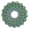
 マップデータ マップデータ | Reconstruction of the immature retroviral CANC hexamer | |||||||||
 試料 試料 |
| |||||||||
 キーワード キーワード | Cryo-electron Tomography / Sub-tomogram Averaging / Retrovirus / Capsid | |||||||||
| 生物種 |  Mason-Pfizer monkey virus (ウイルス) Mason-Pfizer monkey virus (ウイルス) | |||||||||
| 手法 | サブトモグラム平均法 / クライオ電子顕微鏡法 / 解像度: 8.5 Å | |||||||||
 データ登録者 データ登録者 | Schur FKM / Hagen W / de Marco A / Briggs JAG | |||||||||
 引用 引用 |  ジャーナル: J Struct Biol / 年: 2013 ジャーナル: J Struct Biol / 年: 2013タイトル: Determination of protein structure at 8.5Å resolution using cryo-electron tomography and sub-tomogram averaging. 著者: Florian K M Schur / Wim J H Hagen / Alex de Marco / John A G Briggs /  要旨: Cryo-electron tomography combined with image processing by sub-tomogram averaging is unique in its power to resolve the structures of proteins and macromolecular complexes in situ. Limitations of the ...Cryo-electron tomography combined with image processing by sub-tomogram averaging is unique in its power to resolve the structures of proteins and macromolecular complexes in situ. Limitations of the method, including the low signal to noise ratio within individual images from cryo-tomographic datasets and difficulties in determining the defocus at which the data was collected, mean that to date the very best structures obtained by sub-tomogram averaging are limited to a resolution of approximately 15 Å. Here, by optimizing data collection and defocus determination steps, we have determined the structure of assembled Mason-Pfizer monkey virus Gag protein using sub-tomogram averaging to a resolution of 8.5 Å. At this resolution alpha-helices can be directly and clearly visualized. These data demonstrate for the first time that high-resolution structural information can be obtained from cryo-electron tomograms using sub-tomogram averaging. Sub-tomogram averaging has the potential to allow detailed studies of unsolved and biologically relevant structures under biologically relevant conditions. | |||||||||
| 履歴 |
|
- 構造の表示
構造の表示
| ムービー |
 ムービービューア ムービービューア |
|---|---|
| 構造ビューア | EMマップ:  SurfView SurfView Molmil Molmil Jmol/JSmol Jmol/JSmol |
| 添付画像 |
- ダウンロードとリンク
-EMDBアーカイブ
| マップデータ |  emd_2487.map.gz emd_2487.map.gz | 1.9 MB |  EMDBマップデータ形式 EMDBマップデータ形式 | |
|---|---|---|---|---|
| ヘッダ (付随情報) |  emd-2487-v30.xml emd-2487-v30.xml emd-2487.xml emd-2487.xml | 8.4 KB 8.4 KB | 表示 表示 |  EMDBヘッダ EMDBヘッダ |
| 画像 |  emd_2487.tif emd_2487.tif | 796.1 KB | ||
| アーカイブディレクトリ |  http://ftp.pdbj.org/pub/emdb/structures/EMD-2487 http://ftp.pdbj.org/pub/emdb/structures/EMD-2487 ftp://ftp.pdbj.org/pub/emdb/structures/EMD-2487 ftp://ftp.pdbj.org/pub/emdb/structures/EMD-2487 | HTTPS FTP |
-検証レポート
| 文書・要旨 |  emd_2487_validation.pdf.gz emd_2487_validation.pdf.gz | 225.8 KB | 表示 |  EMDB検証レポート EMDB検証レポート |
|---|---|---|---|---|
| 文書・詳細版 |  emd_2487_full_validation.pdf.gz emd_2487_full_validation.pdf.gz | 224.9 KB | 表示 | |
| XML形式データ |  emd_2487_validation.xml.gz emd_2487_validation.xml.gz | 5.5 KB | 表示 | |
| アーカイブディレクトリ |  https://ftp.pdbj.org/pub/emdb/validation_reports/EMD-2487 https://ftp.pdbj.org/pub/emdb/validation_reports/EMD-2487 ftp://ftp.pdbj.org/pub/emdb/validation_reports/EMD-2487 ftp://ftp.pdbj.org/pub/emdb/validation_reports/EMD-2487 | HTTPS FTP |
-関連構造データ
- リンク
リンク
| EMDBのページ |  EMDB (EBI/PDBe) / EMDB (EBI/PDBe) /  EMDataResource EMDataResource |
|---|
- マップ
マップ
| ファイル |  ダウンロード / ファイル: emd_2487.map.gz / 形式: CCP4 / 大きさ: 6.4 MB / タイプ: IMAGE STORED AS FLOATING POINT NUMBER (4 BYTES) ダウンロード / ファイル: emd_2487.map.gz / 形式: CCP4 / 大きさ: 6.4 MB / タイプ: IMAGE STORED AS FLOATING POINT NUMBER (4 BYTES) | ||||||||||||||||||||||||||||||||||||||||||||||||||||||||||||||||||||
|---|---|---|---|---|---|---|---|---|---|---|---|---|---|---|---|---|---|---|---|---|---|---|---|---|---|---|---|---|---|---|---|---|---|---|---|---|---|---|---|---|---|---|---|---|---|---|---|---|---|---|---|---|---|---|---|---|---|---|---|---|---|---|---|---|---|---|---|---|---|
| 注釈 | Reconstruction of the immature retroviral CANC hexamer | ||||||||||||||||||||||||||||||||||||||||||||||||||||||||||||||||||||
| 投影像・断面図 | 画像のコントロール
画像は Spider により作成 | ||||||||||||||||||||||||||||||||||||||||||||||||||||||||||||||||||||
| ボクセルのサイズ | X=Y=Z: 2.025 Å | ||||||||||||||||||||||||||||||||||||||||||||||||||||||||||||||||||||
| 密度 |
| ||||||||||||||||||||||||||||||||||||||||||||||||||||||||||||||||||||
| 対称性 | 空間群: 1 | ||||||||||||||||||||||||||||||||||||||||||||||||||||||||||||||||||||
| 詳細 | EMDB XML:
CCP4マップ ヘッダ情報:
| ||||||||||||||||||||||||||||||||||||||||||||||||||||||||||||||||||||
-添付データ
- 試料の構成要素
-全体 : M-MPV CANC Gag lattice
| 全体 | 名称: M-MPV CANC Gag lattice |
|---|---|
| 要素 |
|
-超分子 #1000: M-MPV CANC Gag lattice
| 超分子 | 名称: M-MPV CANC Gag lattice / タイプ: sample / ID: 1000 / 集合状態: helical / Number unique components: 1 |
|---|
-分子 #1: M-MPV dPro CANC protein
| 分子 | 名称: M-MPV dPro CANC protein / タイプ: protein_or_peptide / ID: 1 / 集合状態: helical / 組換発現: Yes |
|---|---|
| 由来(天然) | 生物種:  Mason-Pfizer monkey virus (ウイルス) Mason-Pfizer monkey virus (ウイルス) |
| 組換発現 | 生物種:  |
-実験情報
-構造解析
| 手法 | クライオ電子顕微鏡法 |
|---|---|
 解析 解析 | サブトモグラム平均法 |
| 試料の集合状態 | helical array |
- 試料調製
試料調製
| 緩衝液 | pH: 7.7 / 詳細: 50mM Tris, 100mM NaCl,1uM Zn |
|---|---|
| グリッド | 詳細: 300mesh copper, glow discharged |
| 凍結 | 凍結剤: ETHANE / 装置: OTHER |
- 電子顕微鏡法
| 顕微鏡 | FEI TITAN KRIOS |
|---|---|
| 特殊光学系 | エネルギーフィルター - 名称: GIF2002 postcolumn energy filter |
| 日付 | 2013年5月21日 |
| 撮影 | カテゴリ: CCD フィルム・検出器のモデル: GENERIC GATAN (2k x 2k) 平均電子線量: 40 e/Å2 |
| 電子線 | 加速電圧: 200 kV / 電子線源:  FIELD EMISSION GUN FIELD EMISSION GUN |
| 電子光学系 | 照射モード: FLOOD BEAM / 撮影モード: BRIGHT FIELD / Cs: 2.7 mm / 最大 デフォーカス(公称値): 3.3 µm / 最小 デフォーカス(公称値): 1.5 µm / 倍率(公称値): 42000 |
| 試料ステージ | 試料ホルダー: Liquid nitrogen cooled 試料ホルダーモデル: FEI TITAN KRIOS AUTOGRID HOLDER Tilt series - Axis1 - Min angle: -45 ° / Tilt series - Axis1 - Max angle: 60 ° |
| 実験機器 |  モデル: Titan Krios / 画像提供: FEI Company |
- 画像解析
画像解析
| 詳細 | Subtomogram averaging was performed using scripts derived from the TOM (Nickell et al, 2005) and AV3 (Foerster et al, 2005) packages. |
|---|---|
| 最終 再構成 | 想定した対称性 - 点群: C2 (2回回転対称) / 解像度のタイプ: BY AUTHOR / 解像度: 8.5 Å / 解像度の算出法: OTHER / ソフトウェア - 名称: TOM, AV3 詳細: Reconstruction carried out using sub-tomogram averaging 使用したサブトモグラム数: 121346 |
| CTF補正 | 詳細: Phase flipping of individual tilts |
 ムービー
ムービー コントローラー


 Z (Sec.)
 Y (Row.)
 X (Col.)


