 Movie
Movie Controller


+ Open data

- Basic information
| Entry | 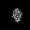 | |||||||||
|---|---|---|---|---|---|---|---|---|---|---|
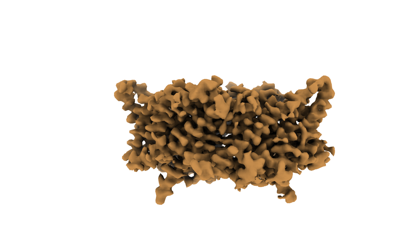
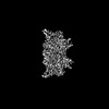
| Title | CLC-ec1 at pH 4.5 100mM Cl Turn | |||||||||
 Map data Map data | CLC-ec1 100 mM Cl pH 4.5 TURN | |||||||||
 Sample Sample |
| |||||||||
 Keywords Keywords | CLC / chloride transporter / MEMBRANE PROTEIN | |||||||||
| Function / homology | Chloride channel, ClcA / Chloride channel, voltage gated / Chloride channel, core / Voltage gated chloride channel / voltage-gated chloride channel activity / antiporter activity / plasma membrane / H(+)/Cl(-) exchange transporter ClcA Function and homology information Function and homology information | |||||||||
| Biological species |  | |||||||||
| Method | single particle reconstruction / cryo EM / Resolution: 3.72 Å | |||||||||
 Authors Authors | Fortea E / Boudker O | |||||||||
| Funding support |  United States, 1 items United States, 1 items
| |||||||||
 Citation Citation | Journal: To Be Published Title: Structural basis of common gate activation in CLC transporters Authors: Fortea E / Lee S / Argyros Y / Chadda R / Ciftci D / Huysmans G / Robertson JL / Boudker O / Accardi A | |||||||||
| History |
|
- Structure visualization
| Supplemental images |
|---|
- Downloads & links
-EMDB archive
| Map data | emd_24612.map.gz | 2.5 MB | EMDB map data format | |
|---|---|---|---|---|
| Header (meta data) | emd-24612-v30.xmlemd-24612.xml | 9.3 KB 9.3 KB | Display Display | EMDB header |
| Images |  emd_24612.png emd_24612.png | 45 KB | ||
| Filedesc metadata | emd-24612.cif.gz | 5.2 KB | ||
| Archive directory |  http://ftp.pdbj.org/pub/emdb/structures/EMD-24612ftp://ftp.pdbj.org/pub/emdb/structures/EMD-24612 http://ftp.pdbj.org/pub/emdb/structures/EMD-24612ftp://ftp.pdbj.org/pub/emdb/structures/EMD-24612 | HTTPS FTP |
-Related structure data
| Related structure data | 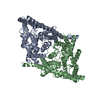 7rp5MC 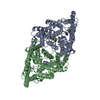 7n8pC 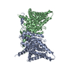 7n9wC 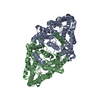 7rnxC 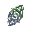 7ro0C 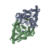 7rp6C 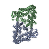 7rq7C 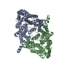 7rsbC M: atomic model generated by this map C: citing same article ( |
|---|---|
| Similar structure data |


-Links
| EMDB pages | EMDB (EBI/PDBe) / EMDataResource |
|---|
-Map
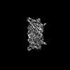
| File | Download / File: emd_24612.map.gz / Format: CCP4 / Size: 28.7 MB / Type: IMAGE STORED AS FLOATING POINT NUMBER (4 BYTES) | ||||||||||||||||||||||||||||||||||||
|---|---|---|---|---|---|---|---|---|---|---|---|---|---|---|---|---|---|---|---|---|---|---|---|---|---|---|---|---|---|---|---|---|---|---|---|---|---|
| Annotation | CLC-ec1 100 mM Cl pH 4.5 TURN | ||||||||||||||||||||||||||||||||||||
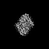
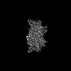
| Projections & slices | Image control
Images are generated by Spider. | ||||||||||||||||||||||||||||||||||||
| Voxel size | X=Y=Z: 1.048 Å | ||||||||||||||||||||||||||||||||||||
| Density |
| ||||||||||||||||||||||||||||||||||||
| Symmetry | Space group: 1 | ||||||||||||||||||||||||||||||||||||
| Details | EMDB XML:
|
 Z (Sec.)
 Y (Row.)
 X (Col.)


-Supplemental data
- Sample components
-Entire : Structure of ecCLC at pH 4.5 in 100mM Cl
| Entire | Name: Structure of ecCLC at pH 4.5 in 100mM Cl |
|---|---|
| Components |
|
-Supramolecule #1: Structure of ecCLC at pH 4.5 in 100mM Cl
| Supramolecule | Name: Structure of ecCLC at pH 4.5 in 100mM Cl / type: complex / ID: 1 / Parent: 0 / Macromolecule list: all |
|---|---|
| Source (natural) | Organism: |
| Molecular weight | Theoretical: 100 kDa/nm |
-Macromolecule #1: H(+)/Cl(-) exchange transporter ClcA
| Macromolecule | Name: H(+)/Cl(-) exchange transporter ClcA / type: protein_or_peptide / ID: 1 / Number of copies: 2 / Enantiomer: LEVO |
|---|---|
| Source (natural) | Organism: |
| Molecular weight | Theoretical: 50.390402 KDa |
| Recombinant expression | Organism: |
| Sequence | String: MKTDTPSLET PQAARLRRRQ LIRQLLERDK TPLAILFMAA VVGTLVGLAA VAFDKGVAWL QNQRMGALVH TADNYPLLLT VAFLCSAVL AMFGYFLVRK YAPEAGGSGI PEIEGALEDQ RPVRWWRVLP VKFFGGLGTL GGGMVLGREG PTVQIGGNIG R MVLDIFRL ...String: MKTDTPSLET PQAARLRRRQ LIRQLLERDK TPLAILFMAA VVGTLVGLAA VAFDKGVAWL QNQRMGALVH TADNYPLLLT VAFLCSAVL AMFGYFLVRK YAPEAGGSGI PEIEGALEDQ RPVRWWRVLP VKFFGGLGTL GGGMVLGREG PTVQIGGNIG R MVLDIFRL KGDEARHTLL ATGAAAGLAA AFNAPLAGIL FIIEEMRPQF RYTLISIKAV FIGVIMSTIM YRIFNHEVAL ID VGKLSDA PLNTLWLYLI LGIIFGIFGP IFNKWVLGMQ DLLHRVHGGN ITKWVLMGGA IGGLCGLLGF VAPATSGGGF NLI PIATAG NFSMGMLVFI FVARVITTLL CFSSGAPGGI FAPMLALGTV LGTAFGMVAV ELFPQYHLEA GTFAIAGMGA LLAA SIRAP LTGIILVLEM TDNYQLILPM IITGLGATLL AQFTGGKPLY SAILARTLAK QEAEQLARSK AASASENT UniProtKB: H(+)/Cl(-) exchange transporter ClcA |
-Experimental details
-Structure determination
| Method | cryo EM |
|---|---|
 Processing Processing | single particle reconstruction |
| Aggregation state | particle |
-Sample preparation
| Concentration | 1.7 mg/mL |
|---|---|
| Buffer | pH: 4.5 |
| Grid | Model: UltrAuFoil R1.2/1.3 |
| Vitrification | Cryogen name: ETHANE / Chamber humidity: 100 % / Chamber temperature: 294.15 K |
- Electron microscopy
| Microscope | FEI TITAN KRIOS |
|---|---|
| Image recording | Film or detector model: GATAN K2 SUMMIT (4k x 4k) / Average electron dose: 72.61 e/Å2 |
| Electron beam | Acceleration voltage: 300 kV / Electron source:  FIELD EMISSION GUN FIELD EMISSION GUN |
| Electron optics | Illumination mode: OTHER / Imaging mode: BRIGHT FIELD / Nominal defocus max: 2.2 µm / Nominal defocus min: 1.2 µm |
| Experimental equipment |  Model: Titan Krios / Image courtesy: FEI Company |
-Image processing
| Startup model | Type of model: NONE |
|---|---|
| Final reconstruction | Resolution.type: BY AUTHOR / Resolution: 3.72 Å / Resolution method: FSC 0.143 CUT-OFF / Number images used: 59069 |
| Initial angle assignment | Type: OTHER |
| Final angle assignment | Type: OTHER |
