Movie
Movie Controller


+ Open data

- Basic information
| Entry | Database: EMDB / ID: EMD-24515 | ||||||||||||
|---|---|---|---|---|---|---|---|---|---|---|---|---|---|
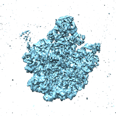
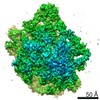
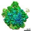
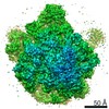
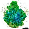
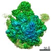
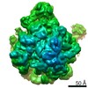
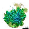
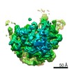
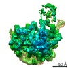
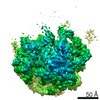
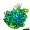
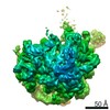
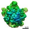
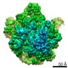
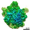
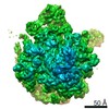
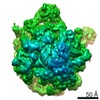
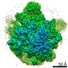
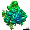
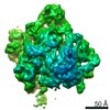
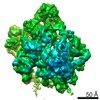
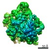
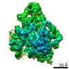
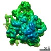
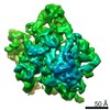
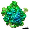
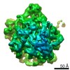
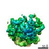
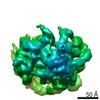
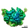
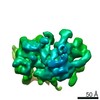
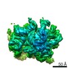
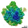
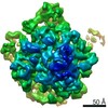
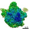
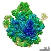
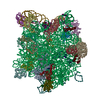
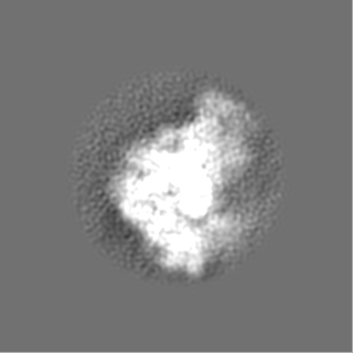
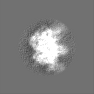
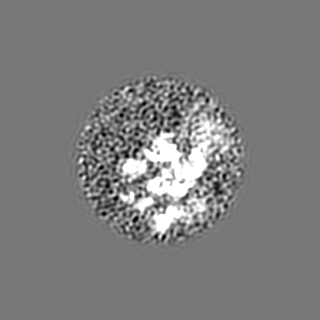
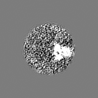
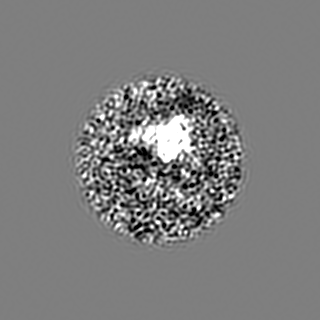
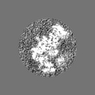
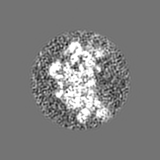
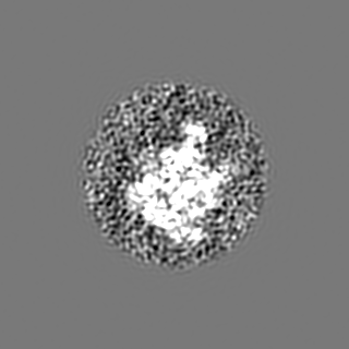
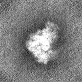
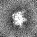
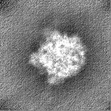
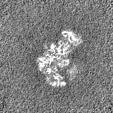
| Title | E. coli bL17-limitation ribosome assembly intermediate #8 | ||||||||||||
 Map data Map data | E. coli bL17-limitation ribosome assembly intermediate #8 (E-class) | ||||||||||||
 Sample Sample |
| ||||||||||||
| Biological species |  | ||||||||||||
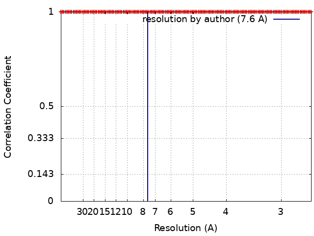
| Method | single particle reconstruction / cryo EM / Resolution: 7.6 Å | ||||||||||||
 Authors Authors | Rabuck-Gibbons JN / Lyumkis D / Williamson JR | ||||||||||||
| Funding support |  United States, 3 items United States, 3 items
| ||||||||||||
 Citation Citation | Journal: Structure / Year: 2022 Title: Quantitative mining of compositional heterogeneity in cryo-EM datasets of ribosome assembly intermediates. Authors: Jessica N Rabuck-Gibbons / Dmitry Lyumkis / James R Williamson / Abstract: Single-particle cryoelectron microscopy (cryo-EM) offers a unique opportunity to characterize macromolecular structural heterogeneity by virtue of its ability to place distinct particle populations ...Single-particle cryoelectron microscopy (cryo-EM) offers a unique opportunity to characterize macromolecular structural heterogeneity by virtue of its ability to place distinct particle populations into different groups through computational classification. However, there is a dearth of tools for surveying the heterogeneity landscape, quantitatively analyzing heterogeneous particle populations after classification, deciding how many unique classes are represented by the data, and accurately cross-comparing reconstructions. Here, we develop a workflow that contains discovery and analysis modules to quantitatively mine cryo-EM data for sets of structures with maximal diversity. This workflow was applied to a dataset of E. coli 50S ribosome assembly intermediates, which are characterized by significant structural heterogeneity. We identified more detailed branchpoints in the assembly process and characterized the interactions of an assembly factor with immature intermediates. While the tools described here were developed for ribosome assembly, they should be broadly applicable to the analysis of other heterogeneous cryo-EM datasets. | ||||||||||||
| History |
|
- Structure visualization
| Movie |
 Movie viewer Movie viewer |
|---|---|
| Structure viewer | EM map: SurfViewMolmilJmol/JSmol |
| Supplemental images |
- Downloads & links
-EMDB archive
| Map data | emd_24515.map.gz | 99.2 MB | EMDB map data format | |
|---|---|---|---|---|
| Header (meta data) | emd-24515-v30.xmlemd-24515.xml | 24.8 KB 24.8 KB | Display Display | EMDB header |
| FSC (resolution estimation) | emd_24515_fsc.xml | 12.2 KB | Display | FSC data file |
| Images |  emd_24515.png emd_24515.png | 147 KB | ||
| Others | emd_24515_additional_1.map.gzemd_24515_additional_2.map.gzemd_24515_additional_3.map.gzemd_24515_half_map_1.map.gzemd_24515_half_map_2.map.gz | 14.5 MB 39.2 KB 16.2 MB 16.3 MB 16.3 MB | ||
| Archive directory |  http://ftp.pdbj.org/pub/emdb/structures/EMD-24515ftp://ftp.pdbj.org/pub/emdb/structures/EMD-24515 http://ftp.pdbj.org/pub/emdb/structures/EMD-24515ftp://ftp.pdbj.org/pub/emdb/structures/EMD-24515 | HTTPS FTP |
-Related structure data
| Related structure data | C: citing same article ( |
|---|---|
| Similar structure data | |
| EM raw data | EMPIAR-10841 (Title: Raw movies and final particle stack for a dataset of bL17-limited E. coli ribosome assembly intermediates. Data size: 1.3 TB Data #1: Particle stack file of L17-depleted ribosomal subunits used for 3D classification [picked particles - single frame - processed] Data #2: Movies of L17-depleted ribosomal subunits used for 3D classification [micrographs - multiframe]) |


-Links
| EMDB pages | EMDB (EBI/PDBe) / EMDataResource |
|---|---|
| Related items in Molecule of the Month |

-Map
| File | Download / File: emd_24515.map.gz / Format: CCP4 / Size: 125 MB / Type: IMAGE STORED AS FLOATING POINT NUMBER (4 BYTES) | ||||||||||||||||||||||||||||||||||||||||||||||||||||||||||||||||||||
|---|---|---|---|---|---|---|---|---|---|---|---|---|---|---|---|---|---|---|---|---|---|---|---|---|---|---|---|---|---|---|---|---|---|---|---|---|---|---|---|---|---|---|---|---|---|---|---|---|---|---|---|---|---|---|---|---|---|---|---|---|---|---|---|---|---|---|---|---|---|
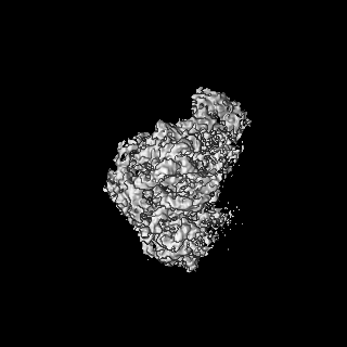
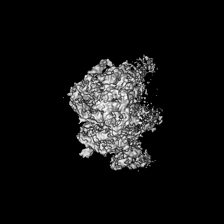
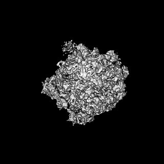
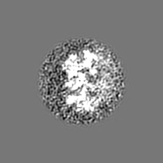
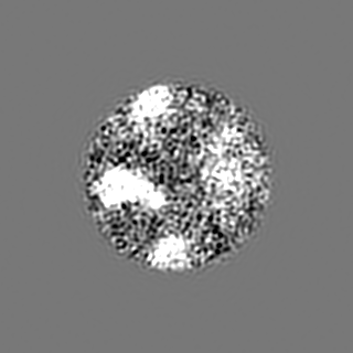
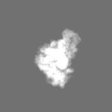
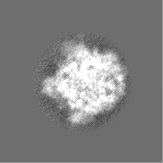
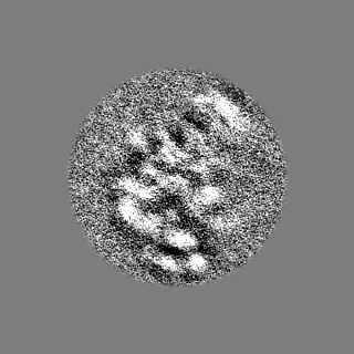
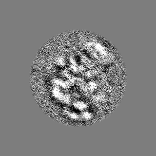
| Annotation | E. coli bL17-limitation ribosome assembly intermediate #8 (E-class) | ||||||||||||||||||||||||||||||||||||||||||||||||||||||||||||||||||||
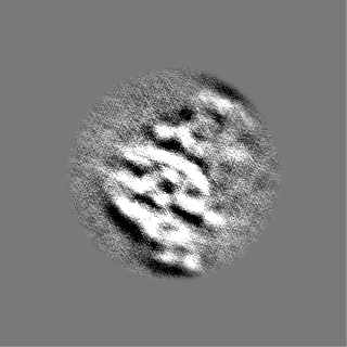
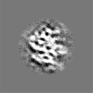
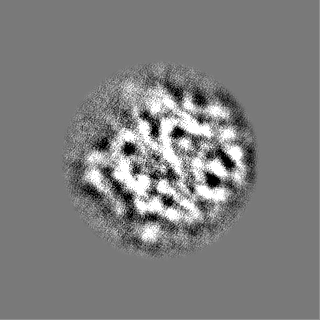
| Projections & slices | Image control
Images are generated by Spider. | ||||||||||||||||||||||||||||||||||||||||||||||||||||||||||||||||||||
| Voxel size | X=Y=Z: 1.31 Å | ||||||||||||||||||||||||||||||||||||||||||||||||||||||||||||||||||||
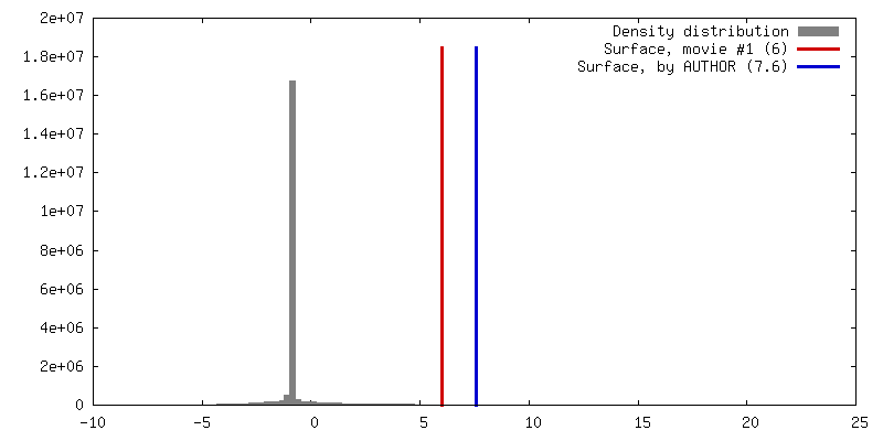
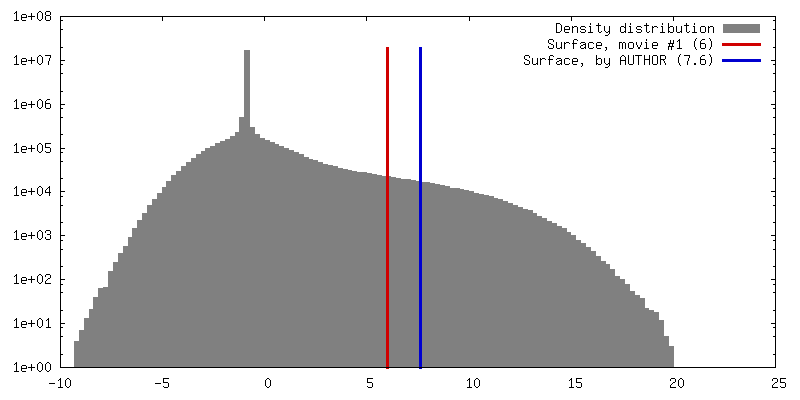
| Density |
| ||||||||||||||||||||||||||||||||||||||||||||||||||||||||||||||||||||
| Symmetry | Space group: 1 | ||||||||||||||||||||||||||||||||||||||||||||||||||||||||||||||||||||
| Details | EMDB XML:
CCP4 map header:
| ||||||||||||||||||||||||||||||||||||||||||||||||||||||||||||||||||||
 Z (Sec.)
 Y (Row.)
 X (Col.)


-Supplemental data
-Additional map: E. coli bL17-limitation ribosome assembly intermediate #8 (E-class)...
| File | emd_24515_additional_1.map | ||||||||||||
|---|---|---|---|---|---|---|---|---|---|---|---|---|---|
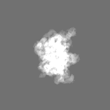
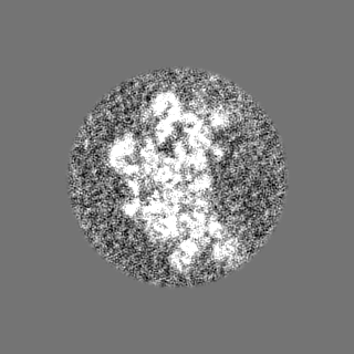
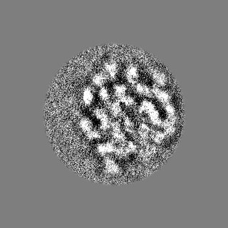
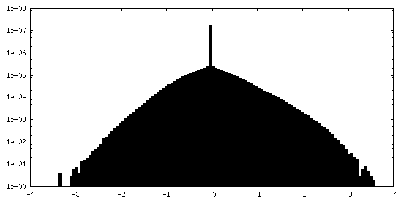
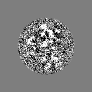
| Annotation | E. coli bL17-limitation ribosome assembly intermediate #8 (E-class) binned, unmasked map | ||||||||||||
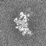
| Projections & Slices |
| ||||||||||||
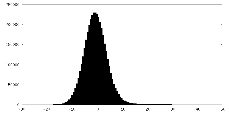
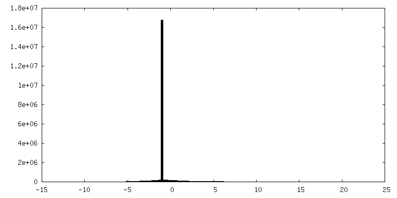
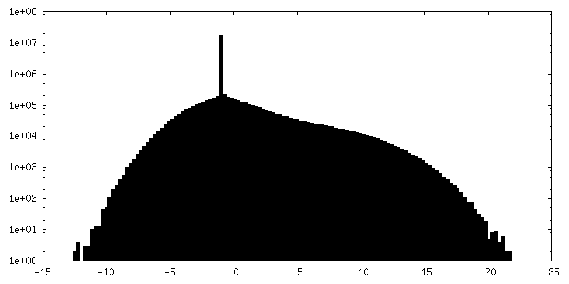
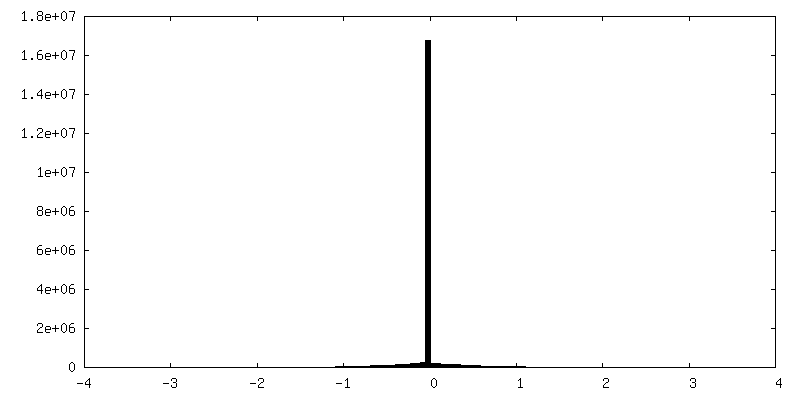
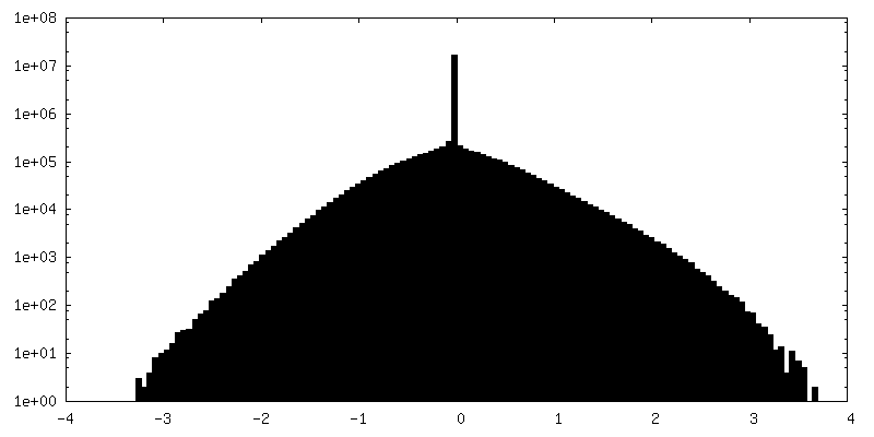
| Density Histograms |
-Additional map: E. coli bL17-limitation ribosome assembly intermediate #8 (E-class)...
| File | emd_24515_additional_2.map | ||||||||||||
|---|---|---|---|---|---|---|---|---|---|---|---|---|---|
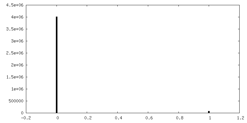
| Annotation | E. coli bL17-limitation ribosome assembly intermediate #8 (E-class) binarized map | ||||||||||||
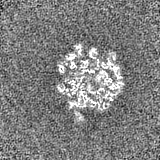
| Projections & Slices |
| ||||||||||||
| Density Histograms |


-Additional map: E. coli bL17-limitation ribosome assembly intermediate #8 (E-class)...
| File | emd_24515_additional_3.map | ||||||||||||
|---|---|---|---|---|---|---|---|---|---|---|---|---|---|
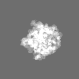
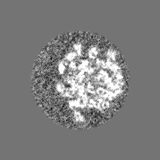
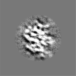
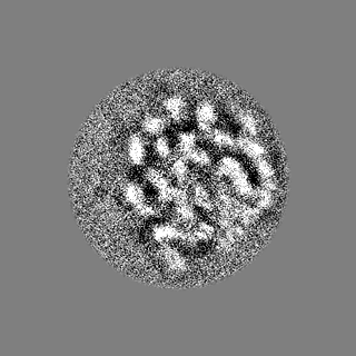
| Annotation | E. coli bL17-limitation ribosome assembly intermediate #8 (E-class) unsharpened map | ||||||||||||
| Projections & Slices |
| ||||||||||||
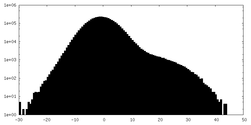
| Density Histograms |


-Half map: E. coli bL17-limitation ribosome assembly intermediate #8 (E-class)...
| File | emd_24515_half_map_1.map | ||||||||||||
|---|---|---|---|---|---|---|---|---|---|---|---|---|---|
| Annotation | E. coli bL17-limitation ribosome assembly intermediate #8 (E-class) odd halfmap | ||||||||||||
| Projections & Slices |
| ||||||||||||
| Density Histograms |


-Half map: E. coli bL17-limitation ribosome assembly intermediate #8 (E-class)...
| File | emd_24515_half_map_2.map | ||||||||||||
|---|---|---|---|---|---|---|---|---|---|---|---|---|---|
| Annotation | E. coli bL17-limitation ribosome assembly intermediate #8 (E-class) even halfmap | ||||||||||||
| Projections & Slices |
| ||||||||||||
| Density Histograms |


- Sample components
-Entire : bL17-depleted large ribosomal subunit assembly intermediate #8
| Entire | Name: bL17-depleted large ribosomal subunit assembly intermediate #8 |
|---|---|
| Components |
|
-Supramolecule #1: bL17-depleted large ribosomal subunit assembly intermediate #8
| Supramolecule | Name: bL17-depleted large ribosomal subunit assembly intermediate #8 type: complex / ID: 1 / Parent: 0 Details: bL17-depleted large ribosomal subunit assembly intermediate #8 |
|---|---|
| Source (natural) | Organism: |
| Molecular weight | Theoretical: 1.5 MDa |
-Experimental details
-Structure determination
| Method | cryo EM |
|---|---|
 Processing Processing | single particle reconstruction |
| Aggregation state | particle |
-Sample preparation
| Buffer | pH: 7.5 Component:
| ||||||||||||
|---|---|---|---|---|---|---|---|---|---|---|---|---|---|
| Grid | Model: Quantifoil R1.2/1.3 / Material: GOLD / Support film - Material: GOLD / Support film - topology: HOLEY | ||||||||||||
| Vitrification | Cryogen name: ETHANE / Instrument: HOMEMADE PLUNGER Details: 3 ul of this sample was added to 3 plasma cleaned (Gatan, Solarus) 1.2mm hole, 1.3mm spacing holey gold grids (Russo and Passmore, 2014). Grids were manually frozen in liquid ethane.. | ||||||||||||
| Details | depleted ribosome assembly intermediate purified by sucrose gradient |
- Electron microscopy
| Microscope | FEI TITAN KRIOS |
|---|---|
| Details | In order to account for highly preferred orientation of the specimen, data was acquired using a tilt of -20 degrees. |
| Image recording | Film or detector model: GATAN K2 SUMMIT (4k x 4k) / Detector mode: SUPER-RESOLUTION / Digitization - Frames/image: 1-50 / Number grids imaged: 1 / Number real images: 833 / Average exposure time: 10.0 sec. / Average electron dose: 35.0 e/Å2 |
| Electron beam | Acceleration voltage: 300 kV / Electron source:  FIELD EMISSION GUN FIELD EMISSION GUN |
| Electron optics | Illumination mode: FLOOD BEAM / Imaging mode: BRIGHT FIELD / Cs: 2.7 mm / Nominal magnification: 22500 |
| Sample stage | Specimen holder model: FEI TITAN KRIOS AUTOGRID HOLDER / Cooling holder cryogen: NITROGEN |
| Experimental equipment |  Model: Titan Krios / Image courtesy: FEI Company |