 ムービー
ムービー コントローラー


+ データを開く

- 基本情報
基本情報
| 登録情報 | データベース: EMDB / ID: EMD-2415 | |||||||||
|---|---|---|---|---|---|---|---|---|---|---|
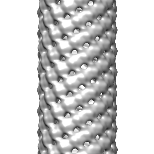
| タイトル | Helical reconstruction of HMPV matrix protein-lipid filaments | |||||||||
 マップデータ マップデータ | Helical reconstruction of human metapneumovirus matrix protein M bound to DOPC | |||||||||
 試料 試料 |
| |||||||||
 キーワード キーワード | human metapneumovirus / HMPV / matrix | |||||||||
| 機能・相同性 |  機能・相同性情報 機能・相同性情報virion assembly / structural constituent of virion / host cell cytoplasm / viral envelope / host cell nucleus / host cell plasma membrane / metal ion binding / identical protein binding 類似検索 - 分子機能 | |||||||||
| 生物種 |  Human metapneumovirus (ウイルス) / synthetic construct (人工物) Human metapneumovirus (ウイルス) / synthetic construct (人工物) | |||||||||
| 手法 | らせん対称体再構成法 / ネガティブ染色法 / 解像度: 28.0 Å | |||||||||
 データ登録者 データ登録者 | Leyrat C / Renner M / Harlos K / Huiskonen JT / Grimes JM | |||||||||
 引用 引用 | ジャーナル: Structure / 年: 2014 タイトル: Structure and self-assembly of the calcium binding matrix protein of human metapneumovirus. 著者: Cedric Leyrat / Max Renner / Karl Harlos / Juha T Huiskonen / Jonathan M Grimes /  要旨: The matrix protein (M) of paramyxoviruses plays a key role in determining virion morphology by directing viral assembly and budding. Here, we report the crystal structure of the human metapneumovirus ...The matrix protein (M) of paramyxoviruses plays a key role in determining virion morphology by directing viral assembly and budding. Here, we report the crystal structure of the human metapneumovirus M at 2.8 Å resolution in its native dimeric state. The structure reveals the presence of a high-affinity Ca²⁺ binding site. Molecular dynamics simulations (MDS) predict a secondary lower-affinity site that correlates well with data from fluorescence-based thermal shift assays. By combining small-angle X-ray scattering with MDS and ensemble analysis, we captured the structure and dynamics of M in solution. Our analysis reveals a large positively charged patch on the protein surface that is involved in membrane interaction. Structural analysis of DOPC-induced polymerization of M into helical filaments using electron microscopy leads to a model of M self-assembly. The conservation of the Ca²⁺ binding sites suggests a role for calcium in the replication and morphogenesis of pneumoviruses. | |||||||||
| 履歴 |
|
- 構造の表示
構造の表示
| ムービー |
ムービービューア |
|---|---|
| 構造ビューア | EMマップ: SurfViewMolmilJmol/JSmol |
| 添付画像 |
- ダウンロードとリンク
-EMDBアーカイブ
| マップデータ | emd_2415.map.gz | 55.6 MB | EMDBマップデータ形式 | |
|---|---|---|---|---|
| ヘッダ (付随情報) | emd-2415-v30.xmlemd-2415.xml | 10.6 KB 10.6 KB | 表示 表示 | EMDBヘッダ |
| 画像 | emd_2415.tif | 148.6 KB | ||
| アーカイブディレクトリ |  http://ftp.pdbj.org/pub/emdb/structures/EMD-2415ftp://ftp.pdbj.org/pub/emdb/structures/EMD-2415 http://ftp.pdbj.org/pub/emdb/structures/EMD-2415ftp://ftp.pdbj.org/pub/emdb/structures/EMD-2415 | HTTPS FTP |
-検証レポート
| 文書・要旨 | emd_2415_validation.pdf.gz | 221.6 KB | 表示 | EMDB検証レポート |
|---|---|---|---|---|
| 文書・詳細版 | emd_2415_full_validation.pdf.gz | 220.7 KB | 表示 | |
| XML形式データ | emd_2415_validation.xml.gz | 6.5 KB | 表示 | |
| アーカイブディレクトリ | https://ftp.pdbj.org/pub/emdb/validation_reports/EMD-2415ftp://ftp.pdbj.org/pub/emdb/validation_reports/EMD-2415 | HTTPS FTP |
-関連構造データ

-リンク
| EMDBのページ | EMDB (EBI/PDBe) / EMDataResource |
|---|
-マップ
| ファイル | ダウンロード / ファイル: emd_2415.map.gz / 形式: CCP4 / 大きさ: 62.5 MB / タイプ: IMAGE STORED AS FLOATING POINT NUMBER (4 BYTES) | ||||||||||||||||||||||||||||||||||||||||||||||||||||||||||||||||||||
|---|---|---|---|---|---|---|---|---|---|---|---|---|---|---|---|---|---|---|---|---|---|---|---|---|---|---|---|---|---|---|---|---|---|---|---|---|---|---|---|---|---|---|---|---|---|---|---|---|---|---|---|---|---|---|---|---|---|---|---|---|---|---|---|---|---|---|---|---|---|
| 注釈 | Helical reconstruction of human metapneumovirus matrix protein M bound to DOPC | ||||||||||||||||||||||||||||||||||||||||||||||||||||||||||||||||||||
| 投影像・断面図 | 画像のコントロール
画像は Spider により作成 | ||||||||||||||||||||||||||||||||||||||||||||||||||||||||||||||||||||
| ボクセルのサイズ | X=Y=Z: 3.1 Å | ||||||||||||||||||||||||||||||||||||||||||||||||||||||||||||||||||||
| 密度 |
| ||||||||||||||||||||||||||||||||||||||||||||||||||||||||||||||||||||
| 対称性 | 空間群: 1 | ||||||||||||||||||||||||||||||||||||||||||||||||||||||||||||||||||||
| 詳細 | EMDB XML:
CCP4マップ ヘッダ情報:
| ||||||||||||||||||||||||||||||||||||||||||||||||||||||||||||||||||||
 Z (Sec.)
 Y (Row.)
 X (Col.)


-添付データ
- 試料の構成要素
-全体 : Human metapneumovirus matrix protein M bound to DOPC
| 全体 | 名称: Human metapneumovirus matrix protein M bound to DOPC |
|---|---|
| 要素 |
|
-超分子 #1000: Human metapneumovirus matrix protein M bound to DOPC
| 超分子 | 名称: Human metapneumovirus matrix protein M bound to DOPC タイプ: sample / ID: 1000 / 集合状態: helical / Number unique components: 2 |
|---|
-分子 #1: human metapneumovirus matrix protein
| 分子 | 名称: human metapneumovirus matrix protein / タイプ: protein_or_peptide / ID: 1 詳細: matrix protein was mixed with DOPC at a final concentration of 0.4 mM 集合状態: dimer / 組換発現: Yes |
|---|---|
| 由来(天然) | 生物種: Human metapneumovirus (ウイルス) / 株: NL1-00 / 別称: Human metapneumovirus |
| 分子量 | 理論値: 55 KDa |
| 組換発現 | 生物種:  |
| 配列 | UniProtKB: Matrix protein / GO: virion assembly, viral envelope / InterPro: Pneumovirus matrix protein |
-分子 #2: 1,2-Dioleoyl-sn-glycero-3-phosphocholine
| 分子 | 名称: 1,2-Dioleoyl-sn-glycero-3-phosphocholine / タイプ: ligand / ID: 2 / Name.synonym: DOPC / 組換発現: No / データベース: NCBI |
|---|---|
| 由来(天然) | 生物種: synthetic construct (人工物) |
| Chemical component information |  ChemComp-PCW: |
-実験情報
-構造解析
| 手法 | ネガティブ染色法 |
|---|---|
 解析 解析 | らせん対称体再構成法 |
| 試料の集合状態 | filament |
-試料調製
| 濃度 | 0.2 mg/mL |
|---|---|
| 緩衝液 | pH: 7.5 / 詳細: 20 mM Tris, 650 mM NaCl, 1M NDSB-201 |
| 染色 | タイプ: NEGATIVE 詳細: Grids with adsorbed protein-lipid mixtures floated on 2% w/v uranyl acetate for 30 seconds |
| グリッド | 詳細: 300 mesh copper grid with formvar carbon film |
| 凍結 | 凍結剤: NONE / 装置: OTHER |
- 電子顕微鏡法
| 顕微鏡 | FEI TECNAI F30 |
|---|---|
| 温度 | 平均: 295 K |
| 日付 | 2013年7月16日 |
| 撮影 | カテゴリ: CCD フィルム・検出器のモデル: GATAN ULTRASCAN 4000 (4k x 4k) デジタル化 - サンプリング間隔: 15 µm / 実像数: 1 / ビット/ピクセル: 12 |
| 電子線 | 加速電圧: 200 kV / 電子線源:  FIELD EMISSION GUN FIELD EMISSION GUN |
| 電子光学系 | 倍率(補正後): 48387 / 照射モード: FLOOD BEAM / 撮影モード: BRIGHT FIELD / 最大 デフォーカス(公称値): 0.82 µm / 最小 デフォーカス(公称値): 0.72 µm / 倍率(公称値): 39000 |
| 試料ステージ | 試料ホルダーモデル: SIDE ENTRY, EUCENTRIC |
| 実験機器 |  モデル: Tecnai F30 / 画像提供: FEI Company |
-画像解析
| 詳細 | The reconstruction was calculated using Burnham-Brandeis Helical Package |
|---|---|
| 最終 再構成 | 想定した対称性 - らせんパラメータ - Δz: 5.16 Å 想定した対称性 - らせんパラメータ - ΔΦ: 56.5 ° アルゴリズム: OTHER / 解像度のタイプ: BY AUTHOR / 解像度: 28.0 Å / 解像度の算出法: OTHER ソフトウェア - 名称: Burnham-Brandeis, Helical, Package 詳細: Final map was filtered to 28 angstrom resolution |
| CTF補正 | 詳細: Each micrograph |
