ムービー
ムービー コントローラー


+ データを開く

- 基本情報
基本情報
| 登録情報 | データベース: EMDB / ID: EMD-24130 | |||||||||
|---|---|---|---|---|---|---|---|---|---|---|
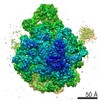
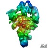
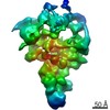
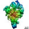
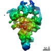
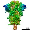
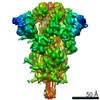
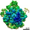
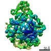
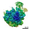
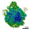
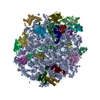
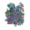
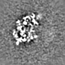
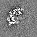
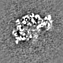
| タイトル | Exploration of subtle structural variability of ribosome assembly intermediates | |||||||||
 マップデータ マップデータ | Exploration of subtle structural variability of ribosome assembly intermediates. | |||||||||
 試料 試料 |
| |||||||||
| 生物種 |  | |||||||||
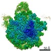
| 手法 | 単粒子再構成法 / クライオ電子顕微鏡法 / 解像度: 8.0 Å | |||||||||
 データ登録者 データ登録者 | Chen M / Ludtke SJ | |||||||||
| 資金援助 |  米国, 1件 米国, 1件
| |||||||||
 引用 引用 | ジャーナル: Nat Methods / 年: 2021 タイトル: Deep learning-based mixed-dimensional Gaussian mixture model for characterizing variability in cryo-EM. 著者: Muyuan Chen / Steven J Ludtke / 要旨: Structural flexibility and/or dynamic interactions with other molecules is a critical aspect of protein function. Cryogenic electron microscopy (cryo-EM) provides direct visualization of individual ...Structural flexibility and/or dynamic interactions with other molecules is a critical aspect of protein function. Cryogenic electron microscopy (cryo-EM) provides direct visualization of individual macromolecules sampling different conformational and compositional states. While numerous methods are available for computational classification of discrete states, characterization of continuous conformational changes or large numbers of discrete state without human supervision remains challenging. Here we present e2gmm, a machine learning algorithm to determine a conformational landscape for proteins or complexes using a three-dimensional Gaussian mixture model mapped onto two-dimensional particle images in known orientations. Using a deep neural network architecture, e2gmm can automatically resolve the structural heterogeneity within the protein complex and map particles onto a small latent space describing conformational and compositional changes. This system presents a more intuitive and flexible representation than other manifold methods currently in use. We demonstrate this method on both simulated data and three biological systems to explore compositional and conformational changes at a range of scales. The software is distributed as part of EMAN2. | |||||||||
| 履歴 |
|
- 構造の表示
構造の表示
| ムービー |
 ムービービューア ムービービューア |
|---|---|
| 構造ビューア | EMマップ: SurfViewMolmilJmol/JSmol |
| 添付画像 |

- ダウンロードとリンク
-EMDBアーカイブ
| マップデータ | emd_24130.map.gz | 7.4 MB | EMDBマップデータ形式 | |
|---|---|---|---|---|
| ヘッダ (付随情報) | emd-24130-v30.xmlemd-24130.xml | 20.7 KB 20.7 KB | 表示 表示 | EMDBヘッダ |
| 画像 | 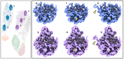 emd_24130.png emd_24130.png | 112.4 KB | ||
| その他 | emd_24130_additional_1.map.gzemd_24130_additional_2.map.gzemd_24130_additional_3.map.gzemd_24130_additional_4.map.gzemd_24130_additional_5.map.gzemd_24130_additional_6.map.gz | 7.4 MB 7.4 MB 7.4 MB 7.4 MB 7.4 MB 7.4 MB | ||
| アーカイブディレクトリ |  http://ftp.pdbj.org/pub/emdb/structures/EMD-24130ftp://ftp.pdbj.org/pub/emdb/structures/EMD-24130 http://ftp.pdbj.org/pub/emdb/structures/EMD-24130ftp://ftp.pdbj.org/pub/emdb/structures/EMD-24130 | HTTPS FTP |
-関連構造データ
| 関連構造データ | C: 同じ文献を引用 ( |
|---|---|
| 類似構造データ |


-リンク
| EMDBのページ | EMDB (EBI/PDBe) / EMDataResource |
|---|---|
| 「今月の分子」の関連する項目 |

-マップ
| ファイル | ダウンロード / ファイル: emd_24130.map.gz / 形式: CCP4 / 大きさ: 8 MB / タイプ: IMAGE STORED AS FLOATING POINT NUMBER (4 BYTES) | ||||||||||||||||||||||||||||||||||||||||||||||||||||||||||||
|---|---|---|---|---|---|---|---|---|---|---|---|---|---|---|---|---|---|---|---|---|---|---|---|---|---|---|---|---|---|---|---|---|---|---|---|---|---|---|---|---|---|---|---|---|---|---|---|---|---|---|---|---|---|---|---|---|---|---|---|---|---|
| 注釈 | Exploration of subtle structural variability of ribosome assembly intermediates. | ||||||||||||||||||||||||||||||||||||||||||||||||||||||||||||
| 投影像・断面図 | 画像のコントロール
画像は Spider により作成 | ||||||||||||||||||||||||||||||||||||||||||||||||||||||||||||
| ボクセルのサイズ | X=Y=Z: 2.62 Å | ||||||||||||||||||||||||||||||||||||||||||||||||||||||||||||
| 密度 |
| ||||||||||||||||||||||||||||||||||||||||||||||||||||||||||||
| 対称性 | 空間群: 1 | ||||||||||||||||||||||||||||||||||||||||||||||||||||||||||||
| 詳細 | EMDB XML:
CCP4マップ ヘッダ情報:
| ||||||||||||||||||||||||||||||||||||||||||||||||||||||||||||
 Z (Sec.)
 Y (Row.)
 X (Col.)


-添付データ
-追加マップ: Exploration of subtle structural variability of ribosome assembly...
| ファイル | emd_24130_additional_1.map | ||||||||||||
|---|---|---|---|---|---|---|---|---|---|---|---|---|---|
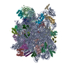
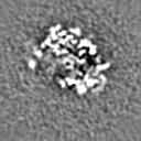
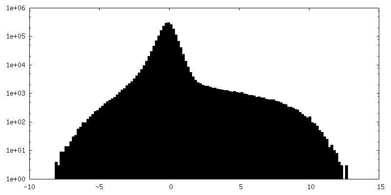
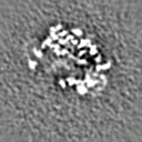
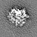
| 注釈 | Exploration of subtle structural variability of ribosome assembly intermediates. Class 2. | ||||||||||||
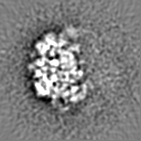
| 投影像・断面図 |
| ||||||||||||
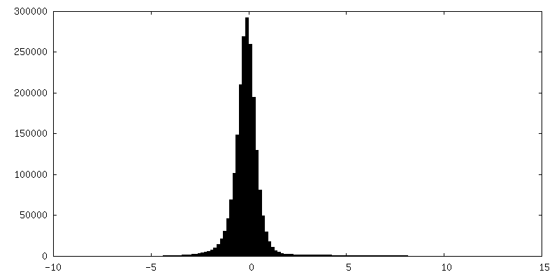
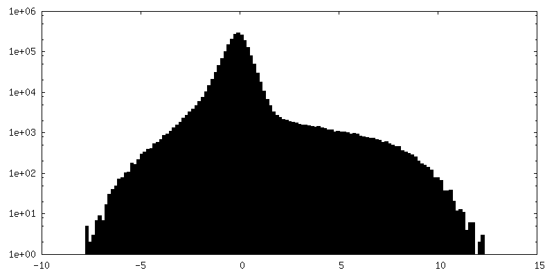
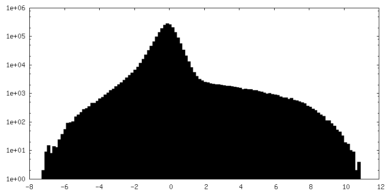
| 密度ヒストグラム |


-追加マップ: Exploration of subtle structural variability of ribosome assembly...
| ファイル | emd_24130_additional_2.map | ||||||||||||
|---|---|---|---|---|---|---|---|---|---|---|---|---|---|
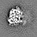
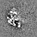
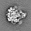
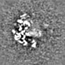
| 注釈 | Exploration of subtle structural variability of ribosome assembly intermediates. Class 1. | ||||||||||||
| 投影像・断面図 |
| ||||||||||||
| 密度ヒストグラム |


-追加マップ: Exploration of subtle structural variability of ribosome assembly...
| ファイル | emd_24130_additional_3.map | ||||||||||||
|---|---|---|---|---|---|---|---|---|---|---|---|---|---|
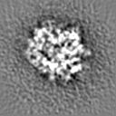
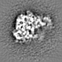
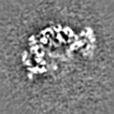
| 注釈 | Exploration of subtle structural variability of ribosome assembly intermediates. Class 0. | ||||||||||||
| 投影像・断面図 |
| ||||||||||||
| 密度ヒストグラム |


-追加マップ: Exploration of subtle structural variability of ribosome assembly...
| ファイル | emd_24130_additional_4.map | ||||||||||||
|---|---|---|---|---|---|---|---|---|---|---|---|---|---|
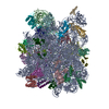
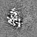
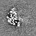
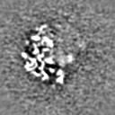
| 注釈 | Exploration of subtle structural variability of ribosome assembly intermediates. Class 5. | ||||||||||||
| 投影像・断面図 |
| ||||||||||||
| 密度ヒストグラム |


-追加マップ: Exploration of subtle structural variability of ribosome assembly...
| ファイル | emd_24130_additional_5.map | ||||||||||||
|---|---|---|---|---|---|---|---|---|---|---|---|---|---|
| 注釈 | Exploration of subtle structural variability of ribosome assembly intermediates. Class 4. | ||||||||||||
| 投影像・断面図 |
| ||||||||||||
| 密度ヒストグラム |


-追加マップ: Exploration of subtle structural variability of ribosome assembly...
| ファイル | emd_24130_additional_6.map | ||||||||||||
|---|---|---|---|---|---|---|---|---|---|---|---|---|---|
| 注釈 | Exploration of subtle structural variability of ribosome assembly intermediates. Class 3. | ||||||||||||
| 投影像・断面図 |
| ||||||||||||
| 密度ヒストグラム |


- 試料の構成要素
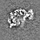
-全体 : bL17-depleted large ribosomal subunit assembly intermediate
| 全体 | 名称: bL17-depleted large ribosomal subunit assembly intermediate |
|---|---|
| 要素 |
|
-超分子 #1: bL17-depleted large ribosomal subunit assembly intermediate
| 超分子 | 名称: bL17-depleted large ribosomal subunit assembly intermediate タイプ: complex / ID: 1 / 親要素: 0 |
|---|---|
| 由来(天然) | 生物種: |
-実験情報
-構造解析
| 手法 | クライオ電子顕微鏡法 |
|---|---|
 解析 解析 | 単粒子再構成法 |
| 試料の集合状態 | particle |
-試料調製
| 緩衝液 | pH: 7 |
|---|---|
| 凍結 | 凍結剤: ETHANE |
- 電子顕微鏡法
| 顕微鏡 | FEI TITAN KRIOS |
|---|---|
| 撮影 | フィルム・検出器のモデル: GATAN K2 SUMMIT (4k x 4k) 平均電子線量: 34.0 e/Å2 |
| 電子線 | 加速電圧: 300 kV / 電子線源:  FIELD EMISSION GUN FIELD EMISSION GUN |
| 電子光学系 | 照射モード: FLOOD BEAM / 撮影モード: BRIGHT FIELD |
| 実験機器 |  モデル: Titan Krios / 画像提供: FEI Company |
-画像解析
| 初期モデル | モデルのタイプ: EMDB MAP EMDB ID: |
|---|---|
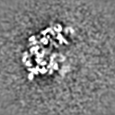
| 最終 再構成 | 使用したクラス数: 6 / アルゴリズム: FOURIER SPACE / 解像度のタイプ: BY AUTHOR / 解像度: 8.0 Å / 解像度の算出法: OTHER 詳細: 2000 particles are included in each class. The maps are filtered to 8A for visualization. 使用した粒子像数: 2000 |
| 初期 角度割当 | タイプ: PROJECTION MATCHING |
| 最終 角度割当 | タイプ: PROJECTION MATCHING / ソフトウェア - 名称: EMAN (ver. 2.91) |