 ムービー
ムービー コントローラー


+ データを開く

- 基本情報
基本情報
| 登録情報 | データベース: EMDB / ID: EMD-2413 | |||||||||
|---|---|---|---|---|---|---|---|---|---|---|
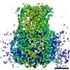
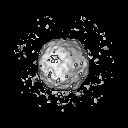
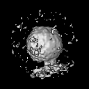
| タイトル | cryo-electron tomography of poliovirus-PVR-liposome complex | |||||||||
 マップデータ マップデータ | Subtomogram asymmetric average of 206 selected virus particles attached to membrane. | |||||||||
 試料 試料 |
| |||||||||
 キーワード キーワード | poliovirus PVR poliovirus receptor liposome 135S | |||||||||
| 生物種 |   Human poliovirus 1 Mahoney (ポリオウイルス) Human poliovirus 1 Mahoney (ポリオウイルス) | |||||||||
| 手法 | サブトモグラム平均法 / クライオ電子顕微鏡法 | |||||||||
 データ登録者 データ登録者 | Strauss M / Levy HC / Bostina M / Filman DJ / Hogle JM | |||||||||
 引用 引用 | ジャーナル: J Virol / 年: 2013 タイトル: RNA transfer from poliovirus 135S particles across membranes is mediated by long umbilical connectors. 著者: Mike Strauss / Hazel C Levy / Mihnea Bostina / David J Filman / James M Hogle /  要旨: During infection, the binding of poliovirus to its cell surface receptor at 37°C triggers an expansion of the virus in which internal polypeptides that bind to membranes are externalized. ...During infection, the binding of poliovirus to its cell surface receptor at 37°C triggers an expansion of the virus in which internal polypeptides that bind to membranes are externalized. Subsequently, in a poorly understood process, the viral RNA genome is transferred directly across an endosomal membrane, and into the host cell cytoplasm, to initiate infection. Here, cryoelectron tomography demonstrates the results of 37°C warming of a poliovirus-receptor-liposome model complex that was produced using Ni-nitrilotriacetic acid lipids and His-tagged receptor ectodomains. In total, 651 subtomographic volumes were aligned, classified, and averaged to obtain detailed pictures, showing both the conversion of virus into its expanded form and the passage of RNA into intact liposomes. Unexpectedly, the virus and membrane surfaces were located ∼50 Å apart, with the 5-fold axis tilted away from the perpendicular, and the solvent spaces between them were spanned by either one or two long "umbilical" density features that lie at an angle to the virus and membrane. The thinner connector, which sometimes appears alone, is 28 to 30 Å in diameter and has a footprint on the virus surface located close to either a 5-fold or a 3-fold axis. The broader connector has a footprint near the quasi-3-fold hole that opens upon virus expansion and is hypothesized to include RNA, shielded from enzymatic degradation by polypeptides that include the N-terminal extension of VP1 and capsid protein VP4. The implications of these observations for the mechanism of RNase-protected RNA transfer in picornaviruses are discussed. | |||||||||
| 履歴 |
|
- 構造の表示
構造の表示
| ムービー |
 ムービービューア ムービービューア |
|---|---|
| 構造ビューア | EMマップ: SurfViewMolmilJmol/JSmol |
| 添付画像 |

- ダウンロードとリンク
-EMDBアーカイブ
| マップデータ | emd_2413.map.gz | 7.1 MB | EMDBマップデータ形式 | |
|---|---|---|---|---|
| ヘッダ (付随情報) | emd-2413-v30.xmlemd-2413.xml | 9 KB 9 KB | 表示 表示 | EMDBヘッダ |
| 画像 |  emd_2413.jpg emd_2413.jpg | 17.4 KB | ||
| アーカイブディレクトリ |  http://ftp.pdbj.org/pub/emdb/structures/EMD-2413ftp://ftp.pdbj.org/pub/emdb/structures/EMD-2413 http://ftp.pdbj.org/pub/emdb/structures/EMD-2413ftp://ftp.pdbj.org/pub/emdb/structures/EMD-2413 | HTTPS FTP |
-検証レポート
| 文書・要旨 | emd_2413_validation.pdf.gz | 229.8 KB | 表示 | EMDB検証レポート |
|---|---|---|---|---|
| 文書・詳細版 | emd_2413_full_validation.pdf.gz | 228.9 KB | 表示 | |
| XML形式データ | emd_2413_validation.xml.gz | 5.8 KB | 表示 | |
| アーカイブディレクトリ | https://ftp.pdbj.org/pub/emdb/validation_reports/EMD-2413ftp://ftp.pdbj.org/pub/emdb/validation_reports/EMD-2413 | HTTPS FTP |
-関連構造データ


-リンク
| EMDBのページ | EMDB (EBI/PDBe) / EMDataResource |
|---|---|
| 「今月の分子」の関連する項目 |

-マップ
| ファイル | ダウンロード / ファイル: emd_2413.map.gz / 形式: CCP4 / 大きさ: 7.8 MB / タイプ: IMAGE STORED AS FLOATING POINT NUMBER (4 BYTES) | ||||||||||||||||||||||||||||||||||||||||||||||||||||||||||||||||||||
|---|---|---|---|---|---|---|---|---|---|---|---|---|---|---|---|---|---|---|---|---|---|---|---|---|---|---|---|---|---|---|---|---|---|---|---|---|---|---|---|---|---|---|---|---|---|---|---|---|---|---|---|---|---|---|---|---|---|---|---|---|---|---|---|---|---|---|---|---|---|
| 注釈 | Subtomogram asymmetric average of 206 selected virus particles attached to membrane. | ||||||||||||||||||||||||||||||||||||||||||||||||||||||||||||||||||||
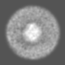
| 投影像・断面図 | 画像のコントロール
画像は Spider により作成 | ||||||||||||||||||||||||||||||||||||||||||||||||||||||||||||||||||||
| ボクセルのサイズ | X=Y=Z: 6.25 Å | ||||||||||||||||||||||||||||||||||||||||||||||||||||||||||||||||||||
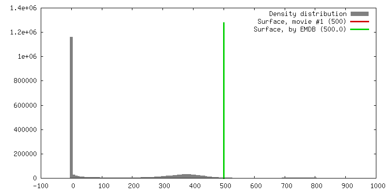
| 密度 |
| ||||||||||||||||||||||||||||||||||||||||||||||||||||||||||||||||||||
| 対称性 | 空間群: 1 | ||||||||||||||||||||||||||||||||||||||||||||||||||||||||||||||||||||
| 詳細 | EMDB XML:
CCP4マップ ヘッダ情報:
| ||||||||||||||||||||||||||||||||||||||||||||||||||||||||||||||||||||
 Z (Sec.)
 Y (Row.)
 X (Col.)


-添付データ
- 試料の構成要素
-全体 : selected "best" average 135S poliovirus - membrane complex
| 全体 | 名称: selected "best" average 135S poliovirus - membrane complex |
|---|---|
| 要素 |
|
-超分子 #1000: selected "best" average 135S poliovirus - membrane complex
| 超分子 | 名称: selected "best" average 135S poliovirus - membrane complex タイプ: sample / ID: 1000 集合状態: one particle connects to one membrane at 2 sites Number unique components: 1 |
|---|
-超分子 #1: Human poliovirus 1 Mahoney
| 超分子 | 名称: Human poliovirus 1 Mahoney / タイプ: virus / ID: 1 詳細: The particle is in the expanded (135S) state, and connected to the membrane. NCBI-ID: 12081 / 生物種: Human poliovirus 1 Mahoney / ウイルスタイプ: VIRION / ウイルス・単離状態: SEROTYPE / ウイルス・エンベロープ: No / ウイルス・中空状態: No / Sci species serotype: PV-1 |
|---|---|
| 宿主 | 生物種:  Homo sapiens (ヒト) / 別称: VERTEBRATES Homo sapiens (ヒト) / 別称: VERTEBRATES |
| 分子量 | 実験値: 8.5 MDa / 理論値: 8.5 MDa |
| ウイルス殻 | Shell ID: 1 / 直径: 330 Å / T番号(三角分割数): 1 |
-実験情報
-構造解析
| 手法 | クライオ電子顕微鏡法 |
|---|---|
 解析 解析 | サブトモグラム平均法 |
| 試料の集合状態 | particle |
-試料調製
| 濃度 | 1 mg/mL |
|---|---|
| 緩衝液 | pH: 7.3 / 詳細: 50mM Hepes, 50mM NaCl |
| グリッド | 詳細: 200 mesh copper Quantifoil grids (R2/2) with 3 nm carbon support on top. |
| 凍結 | 凍結剤: ETHANE / チャンバー内湿度: 90 % / チャンバー内温度: 120 K / 装置: FEI VITROBOT MARK III / 手法: 2 second blot |
- 電子顕微鏡法
| 顕微鏡 | FEI TITAN KRIOS |
|---|---|
| アライメント法 | Legacy - Electron beam tilt params: 0 |
| 日付 | 2010年10月10日 |
| 撮影 | カテゴリ: CCD フィルム・検出器のモデル: GATAN ULTRASCAN 1000 (2k x 2k) 平均電子線量: 60 e/Å2 / ビット/ピクセル: 16 |
| 電子線 | 加速電圧: 300 kV / 電子線源:  FIELD EMISSION GUN FIELD EMISSION GUN |
| 電子光学系 | 倍率(補正後): 45454 / 照射モード: FLOOD BEAM / 撮影モード: BRIGHT FIELD / Cs: 2.7 mm / 倍率(公称値): 50000 |
| 試料ステージ | 試料ホルダーモデル: FEI TITAN KRIOS AUTOGRID HOLDER |
| 実験機器 |  モデル: Titan Krios / 画像提供: FEI Company |
-画像解析
| 詳細 | all particles close to a membrane were averaged |
|---|---|
| 最終 再構成 | 想定した対称性 - 点群: C1 (非対称) ソフトウェア - 名称: IMOD, PEET, BSOFT, EMAN2, SPARX 使用したサブトモグラム数: 206 |
