National Institutes of Health/National Institute Of Allergy and Infectious Diseases (NIH/NIAID)
5R21AI142378-02
United States
Citation
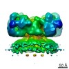
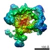
Journal: mBio / Year: 2021 Title: Visualization of the pKM101-Encoded Type IV Secretion System Reveals a Highly Symmetric ATPase Energy Center. Authors: Pratick Khara / Liqiang Song / Peter J Christie / Bo Hu / Abstract: Bacterial conjugation systems are members of the type IV secretion system (T4SS) superfamily. T4SSs can be classified as "minimized" or "expanded" based on whether they are composed of a core set of ...Bacterial conjugation systems are members of the type IV secretion system (T4SS) superfamily. T4SSs can be classified as "minimized" or "expanded" based on whether they are composed of a core set of signature subunits or additional system-specific components. Prototypical minimized systems mediating Agrobacterium tumefaciens transfer DNA (T-DNA) and pKM101 and R388 plasmid transfer are built from subunits generically named VirB1 to VirB11 and VirD4. We visualized the pKM101-encoded T4SS in its native cellular context by cryo-electron tomography (CryoET). The T4SS is composed of an outer membrane core complex (OMCC) connected by a thin stalk to an inner membrane complex (IMC). The OMCC exhibits 14-fold symmetry and resembles that of the T4SS analyzed previously by single-particle electron microscopy. The IMC is highly symmetrical and exhibits 6-fold symmetry. It is dominated by a hexameric collar in the periplasm and a cytoplasmic complex composed of a hexamer of dimers of the VirB4-like TraB ATPase. The IMC closely resembles equivalent regions of three expanded T4SSs previously visualized by CryoET but differs strikingly from the IMC of the purified T4SS, whose cytoplasmic complex instead presents as two side-by-side VirB4 hexamers. Analyses of mutant machines lacking each of the three ATPases required for T4SS function supplied evidence that TraB as well as VirB11-like TraG contribute to distinct stages of machine assembly. We propose that the VirB4-like ATPases, configured as hexamers of dimers at the T4SS entrance, orchestrate IMC assembly and recruitment of the spatially dynamic VirB11 and VirD4 ATPases to activate the T4SS for substrate transfer. Bacterial type IV secretion systems (T4SSs) play central roles in antibiotic resistance spread and virulence. By cryo-electron tomography (CryoET), we solved the structure of the plasmid pKM101-encoded T4SS in the native context of the bacterial cell envelope. The inner membrane complex (IMC) of the T4SS differs remarkably from that of a closely related T4SS analyzed by single-particle electron microscopy. Our findings underscore the importance of comparative and analyses of the T4SS nanomachines and support a unified model in which the signature VirB4 ATPases of the T4SS superfamily function as a central hexamer of dimers to regulate early-stage machine biogenesis and substrate entry passage through the T4SS. The VirB4 ATPases are therefore excellent targets for the development of intervention strategies aimed at suppressing the action of T4SS nanomachines.
History
Deposition
May 24, 2021
-
Header (metadata) release
Dec 15, 2021
-
Map release
Dec 15, 2021
-
Update
Dec 15, 2021
-
Current status
Dec 15, 2021
Processing site: RCSB / Status: Released
-
Structure visualization
Movie
Surface view with section colored by density value
In the structure databanks used in Yorodumi, some data are registered as the other names, "COVID-19 virus" and "2019-nCoV". Here are the details of the virus and the list of structure data.
Jan 31, 2019. EMDB accession codes are about to change! (news from PDBe EMDB page)
EMDB accession codes are about to change! (news from PDBe EMDB page)
The allocation of 4 digits for EMDB accession codes will soon come to an end. Whilst these codes will remain in use, new EMDB accession codes will include an additional digit and will expand incrementally as the available range of codes is exhausted. The current 4-digit format prefixed with “EMD-” (i.e. EMD-XXXX) will advance to a 5-digit format (i.e. EMD-XXXXX), and so on. It is currently estimated that the 4-digit codes will be depleted around Spring 2019, at which point the 5-digit format will come into force.
The EM Navigator/Yorodumi systems omit the EMD- prefix.
Related info.:Q: What is EMD? / ID/Accession-code notation in Yorodumi/EM Navigator
Yorodumi is a browser for structure data from EMDB, PDB, SASBDB, etc.
This page is also the successor to EM Navigator detail page, and also detail information page/front-end page for Omokage search.
The word "yorodu" (or yorozu) is an old Japanese word meaning "ten thousand". "mi" (miru) is to see.
Related info.:EMDB / PDB / SASBDB / Comparison of 3 databanks / Yorodumi Search / Aug 31, 2016. New EM Navigator & Yorodumi / Yorodumi Papers / Jmol/JSmol / Function and homology information / Changes in new EM Navigator and Yorodumi
 Movie
Movie Controller


 Open data
 Basic information
 Map data
 Sample
 Authors
 United States, 1 items
  Citation
 Structure visualization
 Movie viewer
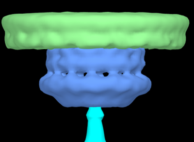
 Downloads & links
 emd_24098.png
 http://ftp.pdbj.org/pub/emdb/structures/EMD-24098

 Z (Sec.)
 Y (Row.)
 X (Col.)


 Sample components
 Processing
 Electron microscopy
 FIELD EMISSION GUN
