Movie
Movie Controller


+ Open data

- Basic information
| Entry |  | |||||||||
|---|---|---|---|---|---|---|---|---|---|---|
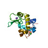
| Title | MicroED structure of lysozyme from milled crystals at 1.75A | |||||||||
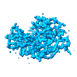
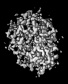
 Map data Map data | 2mFo-Fc map wrapped around protein with 3A carve | |||||||||
 Sample Sample |
| |||||||||
 Keywords Keywords | Lysozyme / HYDROLASE | |||||||||
| Function / homology |  Function and homology information Function and homology informationLactose synthesis / Antimicrobial peptides / Neutrophil degranulation / beta-N-acetylglucosaminidase activity / cell wall macromolecule catabolic process / lysozyme / lysozyme activity / killing of cells of another organism / defense response to Gram-negative bacterium / defense response to bacterium ...Lactose synthesis / Antimicrobial peptides / Neutrophil degranulation / beta-N-acetylglucosaminidase activity / cell wall macromolecule catabolic process / lysozyme / lysozyme activity / killing of cells of another organism / defense response to Gram-negative bacterium / defense response to bacterium / defense response to Gram-positive bacterium / endoplasmic reticulum / extracellular space / identical protein binding / cytoplasm Similarity search - Function | |||||||||
| Biological species |  | |||||||||
| Method | electron crystallography / cryo EM / Resolution: 1.75 Å | |||||||||
 Authors Authors | Martynowycz MW / Gonen T | |||||||||
| Funding support |  United States, 2 items United States, 2 items
| |||||||||
 Citation Citation | Journal: To be Published Title: Preparing crystalline lamellae by focused ion-beam milling for microcrystal electron diffraction (MicroED) experiments Authors: Martynowycz MW / Gonen T | |||||||||
| History |
|
- Structure visualization
| Supplemental images |
|---|
- Downloads & links
-EMDB archive
| Map data | emd_23957.map.gz | 917.4 KB | EMDB map data format | |
|---|---|---|---|---|
| Header (meta data) | emd-23957-v30.xmlemd-23957.xml | 13.4 KB 13.4 KB | Display Display | EMDB header |
| Images |  emd_23957.png emd_23957.png | 53.7 KB | ||
| Filedesc metadata | emd-23957.cif.gz | 5.4 KB | ||
| Filedesc structureFactors | emd_23957_sf.cif.gz | 333.7 KB | ||
| Archive directory |  http://ftp.pdbj.org/pub/emdb/structures/EMD-23957ftp://ftp.pdbj.org/pub/emdb/structures/EMD-23957 http://ftp.pdbj.org/pub/emdb/structures/EMD-23957ftp://ftp.pdbj.org/pub/emdb/structures/EMD-23957 | HTTPS FTP |
-Related structure data
| Related structure data | 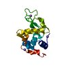 7mrpMC M: atomic model generated by this map C: citing same article ( |
|---|---|
| Similar structure data |
-Links
| EMDB pages | EMDB (EBI/PDBe) / EMDataResource |
|---|---|
| Related items in Molecule of the Month |


-Map
| File | Download / File: emd_23957.map.gz / Format: CCP4 / Size: 4.7 MB / Type: IMAGE STORED AS FLOATING POINT NUMBER (4 BYTES) | ||||||||||||||||||||||||||||||||||||
|---|---|---|---|---|---|---|---|---|---|---|---|---|---|---|---|---|---|---|---|---|---|---|---|---|---|---|---|---|---|---|---|---|---|---|---|---|---|
| Annotation | 2mFo-Fc map wrapped around protein with 3A carve | ||||||||||||||||||||||||||||||||||||
| Projections & slices | Image control
Images are generated by Spider. generated in cubic-lattice coordinate | ||||||||||||||||||||||||||||||||||||
| Voxel size | X: 0.43122 Å / Y: 0.43122 Å / Z: 0.39448 Å | ||||||||||||||||||||||||||||||||||||
| Density |
| ||||||||||||||||||||||||||||||||||||
| Symmetry | Space group: 1 | ||||||||||||||||||||||||||||||||||||
| Details | EMDB XML:
|
 Z (Sec.)
 Y (Row.)
 X (Col.)


-Supplemental data
- Sample components
-Entire : Lysozyme
| Entire | Name: Lysozyme |
|---|---|
| Components |
|
-Supramolecule #1: Lysozyme
| Supramolecule | Name: Lysozyme / type: complex / ID: 1 / Parent: 0 / Macromolecule list: #1 |
|---|---|
| Source (natural) | Organism: |
| Molecular weight | Theoretical: 14.3 KDa |
-Macromolecule #1: Lysozyme C
| Macromolecule | Name: Lysozyme C / type: protein_or_peptide / ID: 1 / Number of copies: 1 / Enantiomer: LEVO / EC number: lysozyme |
|---|---|
| Source (natural) | Organism: |
| Molecular weight | Theoretical: 14.33116 KDa |
| Sequence | String: KVFGRCELAA AMKRHGLDNY RGYSLGNWVC AAKFESNFNT QATNRNTDGS TDYGILQINS RWWCNDGRTP GSRNLCNIPC SALLSSDIT ASVNCAKKIV SDGNGMNAWV AWRNRCKGTD VQAWIRGCRL UniProtKB: Lysozyme C |
-Macromolecule #2: SODIUM ION
| Macromolecule | Name: SODIUM ION / type: ligand / ID: 2 / Number of copies: 4 |
|---|---|
| Molecular weight | Theoretical: 22.99 Da |
-Macromolecule #3: CHLORIDE ION
| Macromolecule | Name: CHLORIDE ION / type: ligand / ID: 3 / Number of copies: 1 / Formula: CL |
|---|---|
| Molecular weight | Theoretical: 35.453 Da |
-Macromolecule #4: water
| Macromolecule | Name: water / type: ligand / ID: 4 / Number of copies: 78 / Formula: HOH |
|---|---|
| Molecular weight | Theoretical: 18.015 Da |
| Chemical component information |  ChemComp-HOH: |
-Experimental details
-Structure determination
| Method | cryo EM |
|---|---|
 Processing Processing | electron crystallography |
| Aggregation state | 3D array |
-Sample preparation
| Concentration | 20 mg/mL |
|---|---|
| Buffer | pH: 4.7 |
| Grid | Model: Quantifoil R2/2 / Material: COPPER / Mesh: 200 / Support film - Material: CARBON / Support film - topology: HOLEY / Support film - Film thickness: 12 / Pretreatment - Type: GLOW DISCHARGE / Pretreatment - Time: 30 sec. |
| Vitrification | Cryogen name: ETHANE / Chamber humidity: 95 % / Chamber temperature: 277 K / Instrument: LEICA EM GP |
- Electron microscopy
| Microscope | TFS TALOS |
|---|---|
| Temperature | Min: 77.0 K / Max: 85.0 K |
| Image recording | Film or detector model: FEI CETA (4k x 4k) / Digitization - Dimensions - Width: 4096 pixel / Digitization - Dimensions - Height: 4096 pixel / Number grids imaged: 1 / Number real images: 1500 / Number diffraction images: 1500 / Average exposure time: 1.0 sec. / Average electron dose: 0.01 e/Å2 |
| Electron beam | Acceleration voltage: 200 kV / Electron source:  FIELD EMISSION GUN FIELD EMISSION GUN |
| Electron optics | C2 aperture diameter: 100.0 µm / Illumination mode: FLOOD BEAM / Imaging mode: DIFFRACTION / Cs: 2.7 mm / Nominal defocus min: 0.0 µm / Camera length: 2100 mm |
| Sample stage | Specimen holder model: FEI TITAN KRIOS AUTOGRID HOLDER / Cooling holder cryogen: NITROGEN / Tilt angle: -30.0, 30.0 |
-Image processing
| Final reconstruction | Algorithm: FOURIER SPACE / Resolution.type: BY AUTHOR / Resolution: 1.75 Å / Resolution method: DIFFRACTION PATTERN/LAYERLINES |
|---|---|
| Crystallography statistics | Number intensities measured: 305349 / Number structure factors: 12144 / Fourier space coverage: 100 / R merge: 0.23 / Overall phase error: 18 / Overall phase residual: 19 / Phase error rejection criteria: none / High resolution: 1.75 Å / Shell - Shell ID: 1 / Shell - High resolution: 1.75 Å / Shell - Low resolution: 34.18 Å / Shell - Number structure factors: 12144 / Shell - Phase residual: 18 / Shell - Fourier space coverage: 100 / Shell - Multiplicity: 25 |