 Movie
Movie Controller


+ Open data

- Basic information
| Entry |  | |||||||||
|---|---|---|---|---|---|---|---|---|---|---|
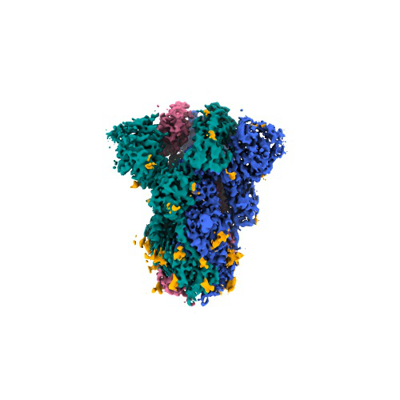
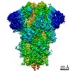
| Title | Prefusion-stabilized SARS-CoV-2 VFLIP_D614G spike | |||||||||
 Map data Map data | ||||||||||
 Sample Sample |
| |||||||||
| Biological species |   Severe acute respiratory syndrome coronavirus 2 Severe acute respiratory syndrome coronavirus 2 | |||||||||
| Method | single particle reconstruction / cryo EM / Resolution: 2.88 Å | |||||||||
 Authors Authors | Olmedillas E / Ollmann Saphire E / Diaz Avalos R | |||||||||
| Funding support |  United States, 1 items United States, 1 items
| |||||||||
 Citation Citation | Journal: To Be Published Title: Prefusion-stabilized SARS-CoV-2 VFLIP_D614G spike Authors: Olmedillas E / Ollmann Saphire E / Diaz Avalos R | |||||||||
| History |
|
- Structure visualization
| Supplemental images |
|---|
- Downloads & links
-EMDB archive
| Map data | emd_23889.map.gz | 229.8 MB |  EMDB map data format EMDB map data format | |
|---|---|---|---|---|
| Header (meta data) | emd-23889-v30.xmlemd-23889.xml | 11.9 KB 11.9 KB | Display Display | EMDB header |
| Images |  emd_23889.png emd_23889.png | 67.1 KB | ||
| Archive directory |  http://ftp.pdbj.org/pub/emdb/structures/EMD-23889ftp://ftp.pdbj.org/pub/emdb/structures/EMD-23889 http://ftp.pdbj.org/pub/emdb/structures/EMD-23889ftp://ftp.pdbj.org/pub/emdb/structures/EMD-23889 | HTTPS FTP |
-Validation report
| Summary document | emd_23889_validation.pdf.gz | 336.7 KB | Display | EMDB validaton report |
|---|---|---|---|---|
| Full document | emd_23889_full_validation.pdf.gz | 336.3 KB | Display | |
| Data in XML | emd_23889_validation.xml.gz | 7 KB | Display | |
| Data in CIF | emd_23889_validation.cif.gz | 8.1 KB | Display | |
| Arichive directory | https://ftp.pdbj.org/pub/emdb/validation_reports/EMD-23889ftp://ftp.pdbj.org/pub/emdb/validation_reports/EMD-23889 | HTTPS FTP |
-Related structure data
| Related structure data |
|---|
-Links
| EMDB pages | EMDB (EBI/PDBe) / EMDataResource |
|---|
-Map
| File | Download / File: emd_23889.map.gz / Format: CCP4 / Size: 244.1 MB / Type: IMAGE STORED AS FLOATING POINT NUMBER (4 BYTES) | ||||||||||||||||||||
|---|---|---|---|---|---|---|---|---|---|---|---|---|---|---|---|---|---|---|---|---|---|
| Voxel size | X=Y=Z: 0.66 Å | ||||||||||||||||||||
| Density |
| ||||||||||||||||||||
| Symmetry | Space group: 1 | ||||||||||||||||||||
| Details | EMDB XML:
|
-Supplemental data
- Sample components
-Entire : Prefusion-stabilized SARS-CoV-2 VFLIP variant D614G spike glycoprotein
| Entire | Name: Prefusion-stabilized SARS-CoV-2 VFLIP variant D614G spike glycoprotein |
|---|---|
| Components |
|
-Supramolecule #1: Prefusion-stabilized SARS-CoV-2 VFLIP variant D614G spike glycoprotein
| Supramolecule | Name: Prefusion-stabilized SARS-CoV-2 VFLIP variant D614G spike glycoprotein type: complex / ID: 1 / Parent: 0 |
|---|---|
| Source (natural) | Organism: Severe acute respiratory syndrome coronavirus 2 |
| Recombinant expression | Organism:   Cricetulus griseus (Chinese hamster) Cricetulus griseus (Chinese hamster) |
| Molecular weight | Theoretical: 660 KDa |
-Experimental details
-Structure determination
| Method | cryo EM |
|---|---|
 Processing Processing | single particle reconstruction |
| Aggregation state | particle |
-Sample preparation
| Concentration | 2 mg/mL | |||||||||
|---|---|---|---|---|---|---|---|---|---|---|
| Buffer | pH: 7.5 Component:
| |||||||||
| Grid | Model: C-flat | |||||||||
| Vitrification | Cryogen name: ETHANE |
- Electron microscopy
| Microscope | FEI TITAN KRIOS |
|---|---|
| Temperature | Min: 83.0 K / Max: 110.0 K |
| Specialist optics | Energy filter - Name: GIF Bioquantum / Energy filter - Slit width: 20 eV |
| Image recording | Film or detector model: GATAN K3 BIOQUANTUM (6k x 4k) / Digitization - Dimensions - Width: 6000 pixel / Digitization - Dimensions - Height: 4000 pixel / Digitization - Sampling interval: 5.0 µm / Number grids imaged: 2 / Number real images: 3000 / Average exposure time: 1.5 sec. / Average electron dose: 50.0 e/Å2 |
| Electron beam | Acceleration voltage: 300 kV / Electron source:  FIELD EMISSION GUN FIELD EMISSION GUN |
| Electron optics | C2 aperture diameter: 70.0 µm / Illumination mode: FLOOD BEAM / Imaging mode: BRIGHT FIELD / Cs: 2.7 mm / Nominal defocus max: 2.0 µm / Nominal defocus min: 0.5 µm / Nominal magnification: 130000 |
| Sample stage | Specimen holder model: FEI TITAN KRIOS AUTOGRID HOLDER / Cooling holder cryogen: NITROGEN |
| Experimental equipment |  Model: Titan Krios / Image courtesy: FEI Company |
+Image processing
-Atomic model buiding 1
| Refinement | Space: REAL / Protocol: RIGID BODY FIT |
|---|
