 ムービー
ムービー コントローラー


+ データを開く

- 基本情報
基本情報
| 登録情報 | データベース: EMDB / ID: EMD-23791 | |||||||||
|---|---|---|---|---|---|---|---|---|---|---|
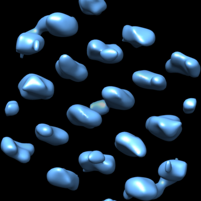
| タイトル | Subtomogram average of Photosystem II 2D semicrystalline array on thylakoid membranes isolated from Phaeodactylum tricornutum | |||||||||
 マップデータ マップデータ | Subtomogram average of Photosystem II 2D semicrystalline array on thylakoid membranes isolated from Phaeodactylum tricornutum | |||||||||
 試料 試料 |
| |||||||||
| 生物種 |  | |||||||||
| 手法 | サブトモグラム平均法 / クライオ電子顕微鏡法 / 解像度: 37.0 Å | |||||||||
 データ登録者 データ登録者 | Jiang J / Cheong KY / Falkowski PG / Dai W | |||||||||
| 資金援助 |  米国, 1件 米国, 1件
| |||||||||
 引用 引用 | ジャーナル: J Struct Biol / 年: 2021 タイトル: Integrating on-grid immunogold labeling and cryo-electron tomography to reveal photosystem II structure and spatial distribution in thylakoid membranes. 著者: Jennifer Jiang / Kuan Yu Cheong / Paul G Falkowski / Wei Dai / 要旨: A long-standing challenge in cell biology is elucidating the structure and spatial distribution of individual membrane-bound proteins, protein complexes and their interactions in their native ...A long-standing challenge in cell biology is elucidating the structure and spatial distribution of individual membrane-bound proteins, protein complexes and their interactions in their native environment. Here, we describe a workflow that combines on-grid immunogold labeling, followed by cryo-electron tomography (cryoET) imaging and structural analyses to identify and characterize the structure of photosystem II (PSII) complexes. Using an antibody specific to a core subunit of PSII, the D1 protein (uniquely found in the water splitting complex in all oxygenic photoautotrophs), we identified PSII complexes in biophysically active thylakoid membranes isolated from a model marine diatom Phaeodactylum tricornutum. Subsequent cryoET analyses of these protein complexes resolved two PSII structures: supercomplexes and dimeric cores. Our integrative approach establishes the structural signature of multimeric membrane protein complexes in their native environment and provides a pathway to elucidate their high-resolution structures. | |||||||||
| 履歴 |
|
- 構造の表示
構造の表示
| ムービー |
 ムービービューア ムービービューア |
|---|---|
| 構造ビューア | EMマップ: SurfViewMolmilJmol/JSmol |
| 添付画像 |
- ダウンロードとリンク
-EMDBアーカイブ
| マップデータ | emd_23791.map.gz | 1.3 MB | EMDBマップデータ形式 | |
|---|---|---|---|---|
| ヘッダ (付随情報) | emd-23791-v30.xmlemd-23791.xml | 10.2 KB 10.2 KB | 表示 表示 | EMDBヘッダ |
| 画像 |  emd_23791.png emd_23791.png | 100.8 KB | ||
| アーカイブディレクトリ |  http://ftp.pdbj.org/pub/emdb/structures/EMD-23791ftp://ftp.pdbj.org/pub/emdb/structures/EMD-23791 http://ftp.pdbj.org/pub/emdb/structures/EMD-23791ftp://ftp.pdbj.org/pub/emdb/structures/EMD-23791 | HTTPS FTP |
-検証レポート
| 文書・要旨 | emd_23791_validation.pdf.gz | 309.7 KB | 表示 | EMDB検証レポート |
|---|---|---|---|---|
| 文書・詳細版 | emd_23791_full_validation.pdf.gz | 309.2 KB | 表示 | |
| XML形式データ | emd_23791_validation.xml.gz | 5.3 KB | 表示 | |
| CIF形式データ | emd_23791_validation.cif.gz | 5.8 KB | 表示 | |
| アーカイブディレクトリ | https://ftp.pdbj.org/pub/emdb/validation_reports/EMD-23791ftp://ftp.pdbj.org/pub/emdb/validation_reports/EMD-23791 | HTTPS FTP |
-関連構造データ
| 類似構造データ |
|---|
-リンク
| EMDBのページ | EMDB (EBI/PDBe) / EMDataResource |
|---|
-マップ
| ファイル | ダウンロード / ファイル: emd_23791.map.gz / 形式: CCP4 / 大きさ: 3.4 MB / タイプ: IMAGE STORED AS FLOATING POINT NUMBER (4 BYTES) | ||||||||||||||||||||||||||||||||||||||||||||||||||||||||||||||||||||
|---|---|---|---|---|---|---|---|---|---|---|---|---|---|---|---|---|---|---|---|---|---|---|---|---|---|---|---|---|---|---|---|---|---|---|---|---|---|---|---|---|---|---|---|---|---|---|---|---|---|---|---|---|---|---|---|---|---|---|---|---|---|---|---|---|---|---|---|---|---|
| 注釈 | Subtomogram average of Photosystem II 2D semicrystalline array on thylakoid membranes isolated from Phaeodactylum tricornutum | ||||||||||||||||||||||||||||||||||||||||||||||||||||||||||||||||||||
| ボクセルのサイズ | X=Y=Z: 5.454 Å | ||||||||||||||||||||||||||||||||||||||||||||||||||||||||||||||||||||
| 密度 |
| ||||||||||||||||||||||||||||||||||||||||||||||||||||||||||||||||||||
| 対称性 | 空間群: 1 | ||||||||||||||||||||||||||||||||||||||||||||||||||||||||||||||||||||
| 詳細 | EMDB XML:
CCP4マップ ヘッダ情報:
| ||||||||||||||||||||||||||||||||||||||||||||||||||||||||||||||||||||
-添付データ
- 試料の構成要素
-全体 : Photosystem II 2D semicrystalline array
| 全体 | 名称: Photosystem II 2D semicrystalline array |
|---|---|
| 要素 |
|
-超分子 #1: Photosystem II 2D semicrystalline array
| 超分子 | 名称: Photosystem II 2D semicrystalline array / タイプ: complex / ID: 1 / 親要素: 0 詳細: On thylakoid membranes isolated from Phaeodactylum tricornutum |
|---|---|
| 由来(天然) | 生物種: |
-実験情報
-構造解析
| 手法 | クライオ電子顕微鏡法 |
|---|---|
 解析 解析 | サブトモグラム平均法 |
| 試料の集合状態 | 2D array |
-試料調製
| 緩衝液 | pH: 7.4 |
|---|---|
| グリッド | モデル: Quantifoil / 材質: COPPER / メッシュ: 200 / 支持フィルム - 材質: CARBON / 支持フィルム - トポロジー: CONTINUOUS / 支持フィルム - Film thickness: 5.0 nm / 前処理 - タイプ: GLOW DISCHARGE |
| 凍結 | 凍結剤: ETHANE / チャンバー内湿度: 95 % / チャンバー内温度: 293.15 K / 装置: LEICA EM GP |
- 電子顕微鏡法
| 顕微鏡 | FEI TALOS ARCTICA |
|---|---|
| 特殊光学系 | エネルギーフィルター - 名称: GIF Bioquantum / エネルギーフィルター - スリット幅: 20 eV |
| 撮影 | フィルム・検出器のモデル: GATAN K2 SUMMIT (4k x 4k) 検出モード: COUNTING / 平均電子線量: 2.0 e/Å2 |
| 電子線 | 加速電圧: 200 kV / 電子線源:  FIELD EMISSION GUN FIELD EMISSION GUN |
| 電子光学系 | C2レンズ絞り径: 100.0 µm / 照射モード: FLOOD BEAM / 撮影モード: BRIGHT FIELD / Cs: 2.7 mm / 倍率(公称値): 49000 |
| 試料ステージ | 試料ホルダーモデル: FEI TITAN KRIOS AUTOGRID HOLDER ホルダー冷却材: NITROGEN |
| 実験機器 |  モデル: Talos Arctica / 画像提供: FEI Company |
-画像解析
| 最終 再構成 | 想定した対称性 - 点群: C2 (2回回転対称) / アルゴリズム: BACK PROJECTION / 解像度のタイプ: BY AUTHOR / 解像度: 37.0 Å / 解像度の算出法: FSC 0.143 CUT-OFF / ソフトウェア - 名称: EMAN2 / 使用したサブトモグラム数: 263 |
|---|---|
| 抽出 | トモグラム数: 1 / 使用した粒子像数: 263 |
| CTF補正 | ソフトウェア - 名称: EMAN2 |
| 最終 角度割当 | タイプ: NOT APPLICABLE |
