 ムービー
ムービー コントローラー


+ データを開く

- 基本情報
基本情報
| 登録情報 | データベース: EMDB / ID: EMD-2374 | |||||||||
|---|---|---|---|---|---|---|---|---|---|---|
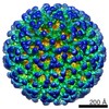
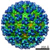
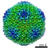
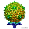
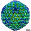
| タイトル | cryo-electron microscopy of Sindbis-liposome complex at pH 6.4 (the group of 5-fold reconstruction) | |||||||||
 マップデータ マップデータ | reconstruction of Sindbis virus when a liposome associates to its 50-fold axis. 5-fold symmetry was applied to the reconstruction map. | |||||||||
 試料 試料 |
| |||||||||
 キーワード キーワード | cryo-electron microscopy / 3D reconstruction / Sindbis virus / membrane fusion / low pH | |||||||||
| 生物種 |  Sindbis virus (シンドビスウイルス) Sindbis virus (シンドビスウイルス) | |||||||||
| 手法 | 単粒子再構成法 / クライオ電子顕微鏡法 / 解像度: 14.5 Å | |||||||||
 データ登録者 データ登録者 | Cao S / Zhang W | |||||||||
 引用 引用 | ジャーナル: Proc Natl Acad Sci U S A / 年: 2013 タイトル: Characterization of an early-stage fusion intermediate of Sindbis virus using cryoelectron microscopy. 著者: Sheng Cao / Wei Zhang /  要旨: The sequential steps in the alphavirus membrane fusion pathway have been postulated based on the prefusion and postfusion crystal structures of the viral fusion protein E1 in conjunction with ...The sequential steps in the alphavirus membrane fusion pathway have been postulated based on the prefusion and postfusion crystal structures of the viral fusion protein E1 in conjunction with biochemical studies. However, the molecular structures of the hypothesized fusion intermediates have remained obscure due to difficulties inherent in the dynamic nature of the process. We developed an experimental system that uses liposomes as the target membrane to capture Sindbis virus, a prototypical alphavirus, in its membrane-binding form at pH 6.4. Cryoelectron micrograph analyses and 3D reconstructions showed that the virus retains its overall icosahedral structure at this mildly acidic pH, except in the membrane-binding region, where monomeric E1 associates with the target membrane and the E2 glycoprotein retains its original trimeric organization. The remaining E2 trimers may hinder E1 homotrimerization and are a potential target for antiviral drugs. | |||||||||
| 履歴 |
|
- 構造の表示
構造の表示
| ムービー |
 ムービービューア ムービービューア |
|---|---|
| 構造ビューア | EMマップ: SurfViewMolmilJmol/JSmol |
| 添付画像 |

- ダウンロードとリンク
-EMDBアーカイブ
| マップデータ | emd_2374.map.gz | 25.3 MB | EMDBマップデータ形式 | |
|---|---|---|---|---|
| ヘッダ (付随情報) | emd-2374-v30.xmlemd-2374.xml | 8.5 KB 8.5 KB | 表示 表示 | EMDBヘッダ |
| 画像 |  emd_2374.png emd_2374.png | 287.8 KB | ||
| アーカイブディレクトリ |  http://ftp.pdbj.org/pub/emdb/structures/EMD-2374ftp://ftp.pdbj.org/pub/emdb/structures/EMD-2374 http://ftp.pdbj.org/pub/emdb/structures/EMD-2374ftp://ftp.pdbj.org/pub/emdb/structures/EMD-2374 | HTTPS FTP |
-関連構造データ
-リンク
| EMDBのページ | EMDB (EBI/PDBe) / EMDataResource |
|---|
-マップ
| ファイル | ダウンロード / ファイル: emd_2374.map.gz / 形式: CCP4 / 大きさ: 61.8 MB / タイプ: IMAGE STORED AS FLOATING POINT NUMBER (4 BYTES) | ||||||||||||||||||||||||||||||||||||||||||||||||||||||||||||||||||||
|---|---|---|---|---|---|---|---|---|---|---|---|---|---|---|---|---|---|---|---|---|---|---|---|---|---|---|---|---|---|---|---|---|---|---|---|---|---|---|---|---|---|---|---|---|---|---|---|---|---|---|---|---|---|---|---|---|---|---|---|---|---|---|---|---|---|---|---|---|---|
| 注釈 | reconstruction of Sindbis virus when a liposome associates to its 50-fold axis. 5-fold symmetry was applied to the reconstruction map. | ||||||||||||||||||||||||||||||||||||||||||||||||||||||||||||||||||||
| 投影像・断面図 | 画像のコントロール
画像は Spider により作成 | ||||||||||||||||||||||||||||||||||||||||||||||||||||||||||||||||||||
| ボクセルのサイズ | X=Y=Z: 3.951 Å | ||||||||||||||||||||||||||||||||||||||||||||||||||||||||||||||||||||
| 密度 |
| ||||||||||||||||||||||||||||||||||||||||||||||||||||||||||||||||||||
| 対称性 | 空間群: 1 | ||||||||||||||||||||||||||||||||||||||||||||||||||||||||||||||||||||
| 詳細 | EMDB XML:
CCP4マップ ヘッダ情報:
| ||||||||||||||||||||||||||||||||||||||||||||||||||||||||||||||||||||
 Z (Sec.)
 Y (Row.)
 X (Col.)


-添付データ
- 試料の構成要素
-全体 : Sindbis virus - liposome complex at pH 6.4
| 全体 | 名称: Sindbis virus - liposome complex at pH 6.4 |
|---|---|
| 要素 |
|
-超分子 #1000: Sindbis virus - liposome complex at pH 6.4
| 超分子 | 名称: Sindbis virus - liposome complex at pH 6.4 / タイプ: sample / ID: 1000 / Number unique components: 1 |
|---|
-超分子 #1: Sindbis virus
| 超分子 | 名称: Sindbis virus / タイプ: virus / ID: 1 詳細: The virus was mixed with liposome and was acidified to pH 6.4 at 37C for 2 min NCBI-ID: 11034 / 生物種: Sindbis virus / Sci species strain: TE12 / ウイルスタイプ: VIRION / ウイルス・単離状態: STRAIN / ウイルス・エンベロープ: Yes / ウイルス・中空状態: No |
|---|---|
| 宿主 | 生物種:  Homo sapiens (ヒト) / 別称: VERTEBRATES Homo sapiens (ヒト) / 別称: VERTEBRATES |
| Host system | 生物種:  Mesocricetus auratus (ネズミ) / 組換細胞: BHK-21 Mesocricetus auratus (ネズミ) / 組換細胞: BHK-21 |
| ウイルス殻 | Shell ID: 1 / 名称: glycoprotein / 直径: 700 Å / T番号(三角分割数): 4 |
-実験情報
-構造解析
| 手法 | クライオ電子顕微鏡法 |
|---|---|
 解析 解析 | 単粒子再構成法 |
| 試料の集合状態 | particle |
-試料調製
| 緩衝液 | pH: 6.4 詳細: Virus and liposome were suspended in TNE (10mM Tris, 200mM NaCl, 1mM EDTA, pH 7.4). The low pH buffer is (50mM MES, 200mM NaCl, pH.6.4) |
|---|---|
| グリッド | 詳細: Ultra-thin carbon film |
| 凍結 | 凍結剤: ETHANE / チャンバー内湿度: 100 % / 装置: FEI VITROBOT MARK III Timed resolved state: Vitrified 2 min after waiting in the chamber (37C) |
- 電子顕微鏡法
| 顕微鏡 | FEI TECNAI F30 |
|---|---|
| 温度 | 平均: 95 K |
| 日付 | 2012年2月4日 |
| 撮影 | カテゴリ: CCD フィルム・検出器のモデル: GATAN ULTRASCAN 4000 (4k x 4k) 平均電子線量: 25 e/Å2 |
| 電子線 | 加速電圧: 300 kV / 電子線源:  FIELD EMISSION GUN FIELD EMISSION GUN |
| 電子光学系 | 倍率(補正後): 75930 / 照射モード: FLOOD BEAM / 撮影モード: BRIGHT FIELD / Cs: 2.0 mm / 最大 デフォーカス(公称値): 4.0 µm / 最小 デフォーカス(公称値): 1.0 µm / 倍率(公称値): 59000 |
| 試料ステージ | 試料ホルダーモデル: GATAN LIQUID NITROGEN / Tilt angle min: 0 / Tilt angle max: 0 |
| 実験機器 |  モデル: Tecnai F30 / 画像提供: FEI Company |
-画像解析
| CTF補正 | 詳細: each particle |
|---|---|
| 最終 再構成 | 想定した対称性 - 点群: C5 (5回回転対称) / アルゴリズム: OTHER / 解像度のタイプ: BY AUTHOR / 解像度: 14.5 Å / 解像度の算出法: FSC 0.5 CUT-OFF / ソフトウェア - 名称: auto3dem / 使用した粒子像数: 196 |
