National Institutes of Health/National Institute of General Medical Sciences (NIH/NIGMS)
1DP2GM123494-01
United States
National Institutes of Health/National Institute of General Medical Sciences (NIH/NIGMS)
5T32GM7240-40
United States
National Science Foundation (NSF, United States)
1920374
United States
National Science Foundation (NSF, United States)
1818344
United States
Citation
Journal: Front Mol Biosci / Year: 2021 Title: Generating Chromosome Geometries in a Minimal Cell From Cryo-Electron Tomograms and Chromosome Conformation Capture Maps. Authors: Benjamin R Gilbert / Zane R Thornburg / Vinson Lam / Fatema-Zahra M Rashid / John I Glass / Elizabeth Villa / Remus T Dame / Zaida Luthey-Schulten / Abstract: JCVI-syn3A is a genetically minimal bacterial cell, consisting of 493 genes and only a single 543 kbp circular chromosome. Syn3A's genome and physical size are approximately one-tenth those of the ...JCVI-syn3A is a genetically minimal bacterial cell, consisting of 493 genes and only a single 543 kbp circular chromosome. Syn3A's genome and physical size are approximately one-tenth those of the model bacterial organism 's, and the corresponding reduction in complexity and scale provides a unique opportunity for whole-cell modeling. Previous work established genome-scale gene essentiality and proteomics data along with its essential metabolic network and a kinetic model of genetic information processing. In addition to that information, whole-cell, spatially-resolved kinetic models require cellular architecture, including spatial distributions of ribosomes and the circular chromosome's configuration. We reconstruct cellular architectures of Syn3A cells at the single-cell level directly from cryo-electron tomograms, including the ribosome distributions. We present a method of generating self-avoiding circular chromosome configurations in a lattice model with a resolution of 11.8 bp per monomer on a 4 nm cubic lattice. Realizations of the chromosome configurations are constrained by the ribosomes and geometry reconstructed from the tomograms and include DNA loops suggested by experimental chromosome conformation capture (3C) maps. Using ensembles of simulated chromosome configurations we predict chromosome contact maps for Syn3A cells at resolutions of 250 bp and greater and compare them to the experimental maps. Additionally, the spatial distributions of ribosomes and the DNA-crowding resulting from the individual chromosome configurations can be used to identify macromolecular structures formed from ribosomes and DNA, such as polysomes and expressomes.
EMPIAR-10686 (Title: Cryo-electron tomography of JCVI-Syn3A (small cell) / Data size: 6.4 Data #1: Unaligned Tilt-series of JCVI-Syn3A (small cell) [tilt series] Data #2: Multi-frame micrographs of each tilt for tilt-series of JCVI-Syn3A (small cell) [micrographs - multiframe])
In the structure databanks used in Yorodumi, some data are registered as the other names, "COVID-19 virus" and "2019-nCoV". Here are the details of the virus and the list of structure data.
Jan 31, 2019. EMDB accession codes are about to change! (news from PDBe EMDB page)
EMDB accession codes are about to change! (news from PDBe EMDB page)
The allocation of 4 digits for EMDB accession codes will soon come to an end. Whilst these codes will remain in use, new EMDB accession codes will include an additional digit and will expand incrementally as the available range of codes is exhausted. The current 4-digit format prefixed with “EMD-” (i.e. EMD-XXXX) will advance to a 5-digit format (i.e. EMD-XXXXX), and so on. It is currently estimated that the 4-digit codes will be depleted around Spring 2019, at which point the 5-digit format will come into force.
The EM Navigator/Yorodumi systems omit the EMD- prefix.
Related info.:Q: What is EMD? / ID/Accession-code notation in Yorodumi/EM Navigator
Yorodumi is a browser for structure data from EMDB, PDB, SASBDB, etc.
This page is also the successor to EM Navigator detail page, and also detail information page/front-end page for Omokage search.
The word "yorodu" (or yorozu) is an old Japanese word meaning "ten thousand". "mi" (miru) is to see.
Related info.:EMDB / PDB / SASBDB / Comparison of 3 databanks / Yorodumi Search / Aug 31, 2016. New EM Navigator & Yorodumi / Yorodumi Papers / Jmol/JSmol / Function and homology information / Changes in new EM Navigator and Yorodumi
 Movie
Movie Controller


 Open data
 Basic information
 Map data
 Sample
 Authors
 United States, 4 items
  Citation
 Structure visualization
 Movie viewer
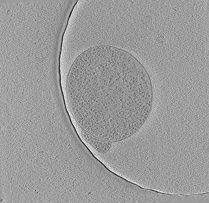
 Downloads & links
 emd_23661.png
 http://ftp.pdbj.org/pub/emdb/structures/EMD-23661
 Sample components
 Processing
 Electron microscopy
 FIELD EMISSION GUN
