National Institutes of Health/National Institute of General Medical Sciences (NIH/NIGMS)
R01GM071940
United States
National Institutes of Health/National Institute Of Allergy and Infectious Diseases (NIH/NIAID)
AI052348
United States
National Institutes of Health/National Institute of General Medical Sciences (NIH/NIGMS)
DE028583
United States
National Institutes of Health/National Center for Research Resources (NIH/NCRR)
S10RR23057
United States
National Institutes of Health/Office of the Director
S10OD018111
United States
National Institutes of Health/National Institute of General Medical Sciences (NIH/NIGMS)
U24GM116792
United States
National Science Foundation (NSF, United States)
DMR-1548924
United States
National Science Foundation (NSF, United States)
DBI-1338135
United States
Swiss National Science Foundation
P300PA_174358
Switzerland
Swiss National Science Foundation
P2BEP3_162094
Switzerland
Citation
Journal: Cell Discov / Year: 2021 Title: Structure of the trypanosome paraflagellar rod and insights into non-planar motility of eukaryotic cells. Authors: Jiayan Zhang / Hui Wang / Simon Imhof / Xueting Zhou / Shiqing Liao / Ivo Atanasov / Wong H Hui / Kent L Hill / Z Hong Zhou / Abstract: Eukaryotic flagella (synonymous with cilia) rely on a microtubule-based axoneme, together with accessory filaments to carryout motility and signaling functions. While axoneme structures are well ...Eukaryotic flagella (synonymous with cilia) rely on a microtubule-based axoneme, together with accessory filaments to carryout motility and signaling functions. While axoneme structures are well characterized, 3D ultrastructure of accessory filaments and their axoneme interface are mostly unknown, presenting a critical gap in understanding structural foundations of eukaryotic flagella. In the flagellum of the protozoan parasite Trypanosoma brucei (T. brucei), the axoneme is accompanied by a paraflagellar rod (PFR) that supports non-planar motility and signaling necessary for disease transmission and pathogenesis. Here, we employed cryogenic electron tomography (cryoET) with sub-tomographic averaging, to obtain structures of the PFR, PFR-axoneme connectors (PACs), and the axonemal central pair complex (CPC). The structures resolve how the 8 nm repeat of the axonemal tubulin dimer interfaces with the 54 nm repeat of the PFR, which consist of proximal, intermediate, and distal zones. In the distal zone, stacked "density scissors" connect with one another to form a "scissors stack network (SSN)" plane oriented 45° to the axoneme axis; and ~370 parallel SSN planes are connected by helix-rich wires into a paracrystalline array with ~90% empty space. Connections from these wires to the intermediate zone, then to overlapping layers of the proximal zone and to the PACs, and ultimately to the CPC, point to a contiguous pathway for signal transmission. Together, our findings provide insights into flagellum-driven, non-planar helical motility of T. brucei and have broad implications ranging from cell motility and tensegrity in biology, to engineering principles in bionics.
History
Deposition
Mar 14, 2021
-
Header (metadata) release
Jul 28, 2021
-
Map release
Jul 28, 2021
-
Update
Jul 28, 2021
-
Current status
Jul 28, 2021
Processing site: RCSB / Status: Released
-
Structure visualization
Movie
Surface view with section colored by density value
In the structure databanks used in Yorodumi, some data are registered as the other names, "COVID-19 virus" and "2019-nCoV". Here are the details of the virus and the list of structure data.
Jan 31, 2019. EMDB accession codes are about to change! (news from PDBe EMDB page)
EMDB accession codes are about to change! (news from PDBe EMDB page)
The allocation of 4 digits for EMDB accession codes will soon come to an end. Whilst these codes will remain in use, new EMDB accession codes will include an additional digit and will expand incrementally as the available range of codes is exhausted. The current 4-digit format prefixed with “EMD-” (i.e. EMD-XXXX) will advance to a 5-digit format (i.e. EMD-XXXXX), and so on. It is currently estimated that the 4-digit codes will be depleted around Spring 2019, at which point the 5-digit format will come into force.
The EM Navigator/Yorodumi systems omit the EMD- prefix.
Related info.:Q: What is EMD? / ID/Accession-code notation in Yorodumi/EM Navigator
Yorodumi is a browser for structure data from EMDB, PDB, SASBDB, etc.
This page is also the successor to EM Navigator detail page, and also detail information page/front-end page for Omokage search.
The word "yorodu" (or yorozu) is an old Japanese word meaning "ten thousand". "mi" (miru) is to see.
Related info.:EMDB / PDB / SASBDB / Comparison of 3 databanks / Yorodumi Search / Aug 31, 2016. New EM Navigator & Yorodumi / Yorodumi Papers / Jmol/JSmol / Function and homology information / Changes in new EM Navigator and Yorodumi
 Movie
Movie Controller

 Yorodumi
 Open data
 Basic information
 Map data
 Sample
 Authors
 United States,
  Switzerland, 10 items
  Citation
 Structure visualization
 Movie viewer
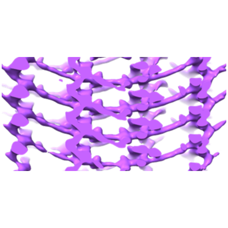
 Downloads & links
 emd_23619.png
 http://ftp.pdbj.org/pub/emdb/structures/EMD-23619
 Z (Sec.)
 Y (Row.)
 X (Col.)


 Sample components
 Processing
 Electron microscopy
 FIELD EMISSION GUN
