 ムービー
ムービー コントローラー


+ データを開く

- 基本情報
基本情報
| 登録情報 | データベース: EMDB / ID: EMD-2328 | |||||||||
|---|---|---|---|---|---|---|---|---|---|---|
| タイトル | Structure of the bacterial V-ATPase from Thermus thermophilius | |||||||||
 マップデータ マップデータ | Reconstruction of the bacterial V-ATPase | |||||||||
 試料 試料 |
| |||||||||
 キーワード キーワード | bacterial V-ATPase / rotary ATPase | |||||||||
| 生物種 |   Thermus thermophilus (バクテリア) Thermus thermophilus (バクテリア) | |||||||||
| 手法 | 電子線結晶学 / ネガティブ染色法 / 解像度: 18.0 Å | |||||||||
 データ登録者 データ登録者 | Tani K / Arthur CP / Tamakoshi M / Yokoyama K / Mitsuoka K / Fujiyoshi Y / Gerle C | |||||||||
 引用 引用 | ジャーナル: Microscopy (Oxf) / 年: 2013 タイトル: Visualization of two distinct states of disassembly in the bacterial V-ATPase from Thermus thermophilus. 著者: Kazutoshi Tani / Christopher P Arthur / Masatada Tamakoshi / Ken Yokoyama / Kaoru Mitsuoka / Yoshinori Fujiyoshi / Christoph Gerle /  要旨: V-ATPases are multisubunit, membrane-bound, energy-converting, cellular machines whose assembly and disassembly is innately connected to their activity in vivo. In vitro V-ATPases show a propensity ...V-ATPases are multisubunit, membrane-bound, energy-converting, cellular machines whose assembly and disassembly is innately connected to their activity in vivo. In vitro V-ATPases show a propensity for disassembly that greatly complicates their functional, and, in particular, structural characterization. Direct structural evidence for early stages of their disassembly has not been reported yet. We analyzed the structure of the V-ATPase from Thermus thermophilus in a single negatively stained two-dimensional (2-D) crystal both by electron tomography and by electron crystallography. Our analysis demonstrated that for 2-D crystals of fragile macromolecular complexes, which are too heterogenous or too few for the merging of image data from many crystals, single-crystal 3-D reconstructions by electron tomography and electron crystallography are expedient tools of analysis. The asymmetric unit in the 2-D crystal lattice contains two different V-ATPase complexes that appear to be in an early stage of disassembly and with either one or both peripheral stalks not being visualized, suggesting the involvement of the peripheral stalks in early stages of disassembly. | |||||||||
| 履歴 |
|
- 構造の表示
構造の表示
| ムービー |
 ムービービューア ムービービューア |
|---|---|
| 構造ビューア | EMマップ: SurfViewMolmilJmol/JSmol |
| 添付画像 |


- ダウンロードとリンク
-EMDBアーカイブ
| マップデータ | emd_2328.map.gz | 424.9 KB | EMDBマップデータ形式 | |
|---|---|---|---|---|
| ヘッダ (付随情報) | emd-2328-v30.xmlemd-2328.xml | 9.5 KB 9.5 KB | 表示 表示 | EMDBヘッダ |
| 画像 | 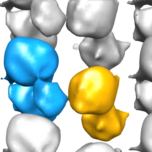 2328_emd-2328-VA_emdep.jpg 2328_emd-2328-VA_emdep.jpg emd-2328-VA_emdep.jpg emd-2328-VA_emdep.jpg | 132.5 KB 132.5 KB | ||
| アーカイブディレクトリ |  http://ftp.pdbj.org/pub/emdb/structures/EMD-2328ftp://ftp.pdbj.org/pub/emdb/structures/EMD-2328 http://ftp.pdbj.org/pub/emdb/structures/EMD-2328ftp://ftp.pdbj.org/pub/emdb/structures/EMD-2328 | HTTPS FTP |
-検証レポート
| 文書・要旨 | emd_2328_validation.pdf.gz | 181.5 KB | 表示 | EMDB検証レポート |
|---|---|---|---|---|
| 文書・詳細版 | emd_2328_full_validation.pdf.gz | 180.6 KB | 表示 | |
| XML形式データ | emd_2328_validation.xml.gz | 4.6 KB | 表示 | |
| アーカイブディレクトリ | https://ftp.pdbj.org/pub/emdb/validation_reports/EMD-2328ftp://ftp.pdbj.org/pub/emdb/validation_reports/EMD-2328 | HTTPS FTP |
-リンク
| EMDBのページ | EMDB (EBI/PDBe) / EMDataResource |
|---|
-マップ
| ファイル | ダウンロード / ファイル: emd_2328.map.gz / 形式: CCP4 / 大きさ: 476.6 KB / タイプ: IMAGE STORED AS FLOATING POINT NUMBER (4 BYTES) | ||||||||||||||||||||||||||||||||||||||||||||||||||||||||||||||||||||
|---|---|---|---|---|---|---|---|---|---|---|---|---|---|---|---|---|---|---|---|---|---|---|---|---|---|---|---|---|---|---|---|---|---|---|---|---|---|---|---|---|---|---|---|---|---|---|---|---|---|---|---|---|---|---|---|---|---|---|---|---|---|---|---|---|---|---|---|---|---|
| 注釈 | Reconstruction of the bacterial V-ATPase | ||||||||||||||||||||||||||||||||||||||||||||||||||||||||||||||||||||
| 投影像・断面図 | 画像のコントロール
画像は Spider により作成 これらの図は立方格子座標系で作成されたものです | ||||||||||||||||||||||||||||||||||||||||||||||||||||||||||||||||||||
| ボクセルのサイズ | X: 4.296 Å / Y: 4.4 Å / Z: 4.166 Å | ||||||||||||||||||||||||||||||||||||||||||||||||||||||||||||||||||||
| 密度 |
| ||||||||||||||||||||||||||||||||||||||||||||||||||||||||||||||||||||
| 対称性 | 空間群: 1 | ||||||||||||||||||||||||||||||||||||||||||||||||||||||||||||||||||||
| 詳細 | EMDB XML:
CCP4マップ ヘッダ情報:
| ||||||||||||||||||||||||||||||||||||||||||||||||||||||||||||||||||||
 Z (Sec.)
 X (Row.)
 Y (Col.)


-添付データ
- 試料の構成要素
-全体 : Bacterial V-type ATPase
| 全体 | 名称: Bacterial V-type ATPase |
|---|---|
| 要素 |
|
-超分子 #1000: Bacterial V-type ATPase
| 超分子 | 名称: Bacterial V-type ATPase / タイプ: sample / ID: 1000 / Number unique components: 1 |
|---|
-分子 #1: V-ATPase
| 分子 | 名称: V-ATPase / タイプ: protein_or_peptide / ID: 1 / コピー数: 2 / 組換発現: No / データベース: NCBI |
|---|---|
| 由来(天然) | 生物種: Thermus thermophilus (バクテリア) / 細胞中の位置: Plasma membrane |
-実験情報
-構造解析
| 手法 | ネガティブ染色法 |
|---|---|
 解析 解析 | 電子線結晶学 |
| 試料の集合状態 | 2D array |
-試料調製
| 濃度 | 1 mg/mL |
|---|---|
| 染色 | タイプ: NEGATIVE 詳細: A 2.5 ul sample was transferred to a glow-discharged carbon coated copper grid. After 1 min, the drop was blotted with a slice of filter paper, and then the grid was washed with 2.5 ul water ...詳細: A 2.5 ul sample was transferred to a glow-discharged carbon coated copper grid. After 1 min, the drop was blotted with a slice of filter paper, and then the grid was washed with 2.5 ul water to avoid the precipitation caused by mixing phosphate buffer with uranyl acetate. Subsequently, the sample was stained with 2.5 ul of 2% uranyl acetate and air-dried. |
| グリッド | 詳細: a glow-discharged carbon coated copper grid |
| 凍結 | 凍結剤: NONE / 装置: OTHER |
| 詳細 | Crystals grown by dialysis |
| 結晶化 | 詳細: Crystals grown by dialysis |
- 電子顕微鏡法
| 顕微鏡 | FEI TECNAI F30 |
|---|---|
| 日付 | 2007年11月28日 |
| 撮影 | カテゴリ: CCD / フィルム・検出器のモデル: GENERIC GATAN / デジタル化 - サンプリング間隔: 15 µm / 実像数: 212 詳細: Every image was recorded by 2Kx2K CCD camera (Gatan) ビット/ピクセル: 16 |
| 電子線 | 加速電圧: 300 kV / 電子線源:  FIELD EMISSION GUN FIELD EMISSION GUN |
| 電子光学系 | 照射モード: FLOOD BEAM / 撮影モード: BRIGHT FIELD / 最大 デフォーカス(公称値): 2.27 µm / 最小 デフォーカス(公称値): 1.4 µm / 倍率(公称値): 40000 |
| 試料ステージ | 試料ホルダーモデル: SIDE ENTRY, EUCENTRIC / Tilt angle min: -55 / Tilt angle max: 55 / Tilt series - Axis1 - Min angle: -55 ° / Tilt series - Axis1 - Max angle: 55 ° |
| 実験機器 |  モデル: Tecnai F30 / 画像提供: FEI Company |
-画像解析
| 詳細 | Images were processed using MRC package |
|---|---|
| 最終 再構成 | 解像度のタイプ: BY AUTHOR / 解像度: 18.0 Å / 解像度の算出法: OTHER / ソフトウェア - 名称: MRC |
| 結晶パラメータ | 単位格子 - A: 232 Å / 単位格子 - B: 132 Å / 単位格子 - C: 300 Å / 単位格子 - γ: 90.0 ° / 単位格子 - α: 90.0 ° / 単位格子 - β: 90.0 ° / 面群: P 1 |
