 Movie
Movie Controller

[English] 日本語
 Yorodumi
- EMDB-23077: Hexameric protein arrangement under docked synaptic vesicles in p... -
+ Open data

- Basic information
| Entry | Database: EMDB / ID: EMD-23077 | |||||||||
|---|---|---|---|---|---|---|---|---|---|---|
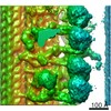
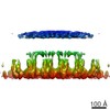
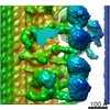
| Title | Hexameric protein arrangement under docked synaptic vesicles in primary hippocampal neurons | |||||||||
 Map data Map data | Cryo-ET/ Subtomogram averaging of protein organization under docked synaptic vesicles from primary hippocampal neurons | |||||||||
 Sample Sample |
| |||||||||
| Biological species |  | |||||||||
| Method | subtomogram averaging / cryo EM / Resolution: 44.0 Å | |||||||||
 Authors Authors | Radhakrishnan A / Li X / Liu J | |||||||||
| Funding support |  United States, 1 items United States, 1 items
| |||||||||
 Citation Citation | Journal: Proc Natl Acad Sci U S A / Year: 2021 Title: Symmetrical arrangement of proteins under release-ready vesicles in presynaptic terminals. Authors: Abhijith Radhakrishnan / Xia Li / Kirill Grushin / Shyam S Krishnakumar / Jun Liu / James E Rothman / Abstract: Controlled release of neurotransmitters stored in synaptic vesicles (SVs) is a fundamental process that is central to all information processing in the brain. This relies on tight coupling of the SV ...Controlled release of neurotransmitters stored in synaptic vesicles (SVs) is a fundamental process that is central to all information processing in the brain. This relies on tight coupling of the SV fusion to action potential-evoked presynaptic Ca influx. This Ca-evoked release occurs from a readily releasable pool (RRP) of SVs docked to the plasma membrane (PM). The protein components involved in initial SV docking/tethering and the subsequent priming reactions which make the SV release ready are known. Yet, the supramolecular architecture and sequence of molecular events underlying SV release are unclear. Here, we use cryoelectron tomography analysis in cultured hippocampal neurons to delineate the arrangement of the exocytosis machinery under docked SVs. Under native conditions, we find that vesicles are initially "tethered" to the PM by a variable number of protein densities (∼10 to 20 nm long) with no discernible organization. In contrast, we observe exactly six protein masses, each likely consisting of a single SNAREpin with its bound Synaptotagmins and Complexin, arranged symmetrically connecting the "primed" vesicles to the PM. Our data indicate that the fusion machinery is likely organized into a highly cooperative framework during the priming process which enables rapid SV fusion and neurotransmitter release following Ca influx. | |||||||||
| History |
|
- Structure visualization
| Movie |
 Movie viewer Movie viewer |
|---|---|
| Structure viewer | EM map: SurfViewMolmilJmol/JSmol |
| Supplemental images |

- Downloads & links
-EMDB archive
| Map data | emd_23077.map.gz | 1.1 MB | EMDB map data format | |
|---|---|---|---|---|
| Header (meta data) | emd-23077-v30.xmlemd-23077.xml | 9 KB 9 KB | Display Display | EMDB header |
| FSC (resolution estimation) | emd_23077_fsc.xml | 3.4 KB | Display | FSC data file |
| Images |  emd_23077.png emd_23077.png | 38.1 KB | ||
| Archive directory |  http://ftp.pdbj.org/pub/emdb/structures/EMD-23077ftp://ftp.pdbj.org/pub/emdb/structures/EMD-23077 http://ftp.pdbj.org/pub/emdb/structures/EMD-23077ftp://ftp.pdbj.org/pub/emdb/structures/EMD-23077 | HTTPS FTP |
-Related structure data
| Similar structure data |
|---|
-Links
| EMDB pages | EMDB (EBI/PDBe) / EMDataResource |
|---|
-Map
| File | Download / File: emd_23077.map.gz / Format: CCP4 / Size: 1.4 MB / Type: IMAGE STORED AS FLOATING POINT NUMBER (4 BYTES) | ||||||||||||||||||||||||||||||||||||||||||||||||||||||||||||
|---|---|---|---|---|---|---|---|---|---|---|---|---|---|---|---|---|---|---|---|---|---|---|---|---|---|---|---|---|---|---|---|---|---|---|---|---|---|---|---|---|---|---|---|---|---|---|---|---|---|---|---|---|---|---|---|---|---|---|---|---|---|
| Annotation | Cryo-ET/ Subtomogram averaging of protein organization under docked synaptic vesicles from primary hippocampal neurons | ||||||||||||||||||||||||||||||||||||||||||||||||||||||||||||
| Projections & slices | Image control
Images are generated by Spider. | ||||||||||||||||||||||||||||||||||||||||||||||||||||||||||||
| Voxel size | X=Y=Z: 10.8 Å | ||||||||||||||||||||||||||||||||||||||||||||||||||||||||||||
| Density |
| ||||||||||||||||||||||||||||||||||||||||||||||||||||||||||||
| Symmetry | Space group: 1 | ||||||||||||||||||||||||||||||||||||||||||||||||||||||||||||
| Details | EMDB XML:
CCP4 map header:
| ||||||||||||||||||||||||||||||||||||||||||||||||||||||||||||
 Z (Sec.)
 Y (Row.)
 X (Col.)


-Supplemental data
- Sample components
-Entire : Docked synaptic vesicles in primary hippocampal neurons
| Entire | Name: Docked synaptic vesicles in primary hippocampal neurons |
|---|---|
| Components |
|
-Supramolecule #1: Docked synaptic vesicles in primary hippocampal neurons
| Supramolecule | Name: Docked synaptic vesicles in primary hippocampal neurons type: organelle_or_cellular_component / ID: 1 / Parent: 0 Details: Primary hippocampal neurons were cultured on EM grids |
|---|---|
| Source (natural) | Organism: |
-Experimental details
-Structure determination
| Method | cryo EM |
|---|---|
 Processing Processing | subtomogram averaging |
| Aggregation state | cell |
-Sample preparation
| Buffer | pH: 7.4 |
|---|---|
| Grid | Model: Quantifoil R2/1 / Material: GOLD / Mesh: 200 / Support film - Material: CARBON / Support film - topology: HOLEY / Pretreatment - Type: GLOW DISCHARGE / Pretreatment - Atmosphere: AIR / Details: 15 mA current |
| Vitrification | Cryogen name: ETHANE / Instrument: HOMEMADE PLUNGER |
| Details | primary hippocampal neurons were cultured directly on EM grids |
- Electron microscopy
| Microscope | FEI TITAN KRIOS |
|---|---|
| Specialist optics | Phase plate: VOLTA PHASE PLATE |
| Image recording | Film or detector model: GATAN K2 SUMMIT (4k x 4k) / Average electron dose: 1.47 e/Å2 |
| Electron beam | Acceleration voltage: 300 kV / Electron source:  FIELD EMISSION GUN FIELD EMISSION GUN |
| Electron optics | Illumination mode: FLOOD BEAM / Imaging mode: BRIGHT FIELD |
| Experimental equipment |  Model: Titan Krios / Image courtesy: FEI Company |
-Image processing
| Final reconstruction | Applied symmetry - Point group: C6 (6 fold cyclic) / Resolution.type: BY AUTHOR / Resolution: 44.0 Å / Resolution method: FSC 0.5 CUT-OFF / Details: Subtomogram averaging / Number subtomograms used: 5726 |
|---|---|
| Extraction | Number tomograms: 296 / Number images used: 7527 |
| Final angle assignment | Type: NOT APPLICABLE |
| FSC plot (resolution estimation) |  |
