National Institutes of Health/National Institute Of Allergy and Infectious Diseases (NIH/NIAID)
75N93019C00074
United States
Citation
Journal: Nature / Year: 2020 Title: Potently neutralizing and protective human antibodies against SARS-CoV-2. Authors: Seth J Zost / Pavlo Gilchuk / James Brett Case / Elad Binshtein / Rita E Chen / Joseph P Nkolola / Alexandra Schäfer / Joseph X Reidy / Andrew Trivette / Rachel S Nargi / Rachel E Sutton / ...Authors: Seth J Zost / Pavlo Gilchuk / James Brett Case / Elad Binshtein / Rita E Chen / Joseph P Nkolola / Alexandra Schäfer / Joseph X Reidy / Andrew Trivette / Rachel S Nargi / Rachel E Sutton / Naveenchandra Suryadevara / David R Martinez / Lauren E Williamson / Elaine C Chen / Taylor Jones / Samuel Day / Luke Myers / Ahmed O Hassan / Natasha M Kafai / Emma S Winkler / Julie M Fox / Swathi Shrihari / Benjamin K Mueller / Jens Meiler / Abishek Chandrashekar / Noe B Mercado / James J Steinhardt / Kuishu Ren / Yueh-Ming Loo / Nicole L Kallewaard / Broc T McCune / Shamus P Keeler / Michael J Holtzman / Dan H Barouch / Lisa E Gralinski / Ralph S Baric / Larissa B Thackray / Michael S Diamond / Robert H Carnahan / James E Crowe / Abstract: The ongoing pandemic of coronavirus disease 2019 (COVID-19), which is caused by severe acute respiratory syndrome coronavirus 2 (SARS-CoV-2), is a major threat to global health and the medical ...The ongoing pandemic of coronavirus disease 2019 (COVID-19), which is caused by severe acute respiratory syndrome coronavirus 2 (SARS-CoV-2), is a major threat to global health and the medical countermeasures available so far are limited. Moreover, we currently lack a thorough understanding of the mechanisms of humoral immunity to SARS-CoV-2. Here we analyse a large panel of human monoclonal antibodies that target the spike (S) glycoprotein, and identify several that exhibit potent neutralizing activity and fully block the receptor-binding domain of the S protein (S) from interacting with human angiotensin-converting enzyme 2 (ACE2). Using competition-binding, structural and functional studies, we show that the monoclonal antibodies can be clustered into classes that recognize distinct epitopes on the S, as well as distinct conformational states of the S trimer. Two potently neutralizing monoclonal antibodies, COV2-2196 and COV2-2130, which recognize non-overlapping sites, bound simultaneously to the S protein and neutralized wild-type SARS-CoV-2 virus in a synergistic manner. In two mouse models of SARS-CoV-2 infection, passive transfer of COV2-2196, COV2-2130 or a combination of both of these antibodies protected mice from weight loss and reduced the viral burden and levels of inflammation in the lungs. In addition, passive transfer of either of two of the most potent ACE2-blocking monoclonal antibodies (COV2-2196 or COV2-2381) as monotherapy protected rhesus macaques from SARS-CoV-2 infection. These results identify protective epitopes on the S and provide a structure-based framework for rational vaccine design and the selection of robust immunotherapeutic agents.
History
Deposition
May 14, 2020
-
Header (metadata) release
May 27, 2020
-
Map release
May 27, 2020
-
Update
Sep 2, 2020
-
Current status
Sep 2, 2020
Processing site: RCSB / Status: Released
-
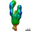
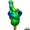
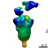
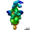
Structure visualization
Movie
Surface view with section colored by density value
In the structure databanks used in Yorodumi, some data are registered as the other names, "COVID-19 virus" and "2019-nCoV". Here are the details of the virus and the list of structure data.
Jan 31, 2019. EMDB accession codes are about to change! (news from PDBe EMDB page)
EMDB accession codes are about to change! (news from PDBe EMDB page)
The allocation of 4 digits for EMDB accession codes will soon come to an end. Whilst these codes will remain in use, new EMDB accession codes will include an additional digit and will expand incrementally as the available range of codes is exhausted. The current 4-digit format prefixed with “EMD-” (i.e. EMD-XXXX) will advance to a 5-digit format (i.e. EMD-XXXXX), and so on. It is currently estimated that the 4-digit codes will be depleted around Spring 2019, at which point the 5-digit format will come into force.
The EM Navigator/Yorodumi systems omit the EMD- prefix.
Related info.:Q: What is EMD? / ID/Accession-code notation in Yorodumi/EM Navigator
Yorodumi is a browser for structure data from EMDB, PDB, SASBDB, etc.
This page is also the successor to EM Navigator detail page, and also detail information page/front-end page for Omokage search.
The word "yorodu" (or yorozu) is an old Japanese word meaning "ten thousand". "mi" (miru) is to see.
Related info.:EMDB / PDB / SASBDB / Comparison of 3 databanks / Yorodumi Search / Aug 31, 2016. New EM Navigator & Yorodumi / Yorodumi Papers / Jmol/JSmol / Function and homology information / Changes in new EM Navigator and Yorodumi
 Movie
Movie Controller

 Yorodumi
 Open data
 Basic information
 Map data
 Sample
 Function and homology information
 Severe acute respiratory syndrome coronavirus 2 /
  Homo sapiens (human)
 Authors
 United States, 2 items
  Citation
 Structure visualization
 Downloads & links
 emd_21975.png
 http://ftp.pdbj.org/pub/emdb/structures/EMD-21975
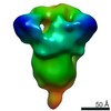


 Z (Sec.)
 Y (Row.)
 X (Col.)


 Sample components
 Processing
 Electron microscopy
 FIELD EMISSION GUN
