 ムービー
ムービー コントローラー


+ データを開く

- 基本情報
基本情報
| 登録情報 | データベース: EMDB / ID: EMD-2186 | |||||||||
|---|---|---|---|---|---|---|---|---|---|---|
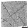
| タイトル | Dual-axis cryo-electron tomogram of microtubules assembled in vitro | |||||||||
 マップデータ マップデータ | Dual-axis cryo-electron tomogram of microtubules assembled in vitro | |||||||||
 試料 試料 |
| |||||||||
 キーワード キーワード | microtubules / single-axis / dual-axis / cryo-electron tomography / missing wedge | |||||||||
| 生物種 |  | |||||||||
| 手法 | 電子線トモグラフィー法 / クライオ電子顕微鏡法 / 解像度: 40.0 Å | |||||||||
 データ登録者 データ登録者 | Guesdon A / Blestel S / Kervrann C / Chretien D | |||||||||
 引用 引用 | ジャーナル: J Struct Biol / 年: 2013 タイトル: Single versus dual-axis cryo-electron tomography of microtubules assembled in vitro: limits and perspectives. 著者: Audrey Guesdon / Sophie Blestel / Charles Kervrann / Denis Chrétien /  要旨: Single-axis cryo-electron tomography of vitrified specimens has become a method of choice to reconstruct in three dimensions macromolecular assemblies in their cellular context or prepared from ...Single-axis cryo-electron tomography of vitrified specimens has become a method of choice to reconstruct in three dimensions macromolecular assemblies in their cellular context or prepared from purified components. Here, we asked how a dual-axis acquisition scheme would improve three-dimensional reconstructions of microtubules assembled in vitro. We show that in single-axis tomograms, microtubules oriented close to the perpendicular of the tilt axis display diminished contrast, and ultimately transform into sets of parallel lines oriented in the direction of the electron beam when observed in cross-section. Analysis of their three-dimensional Fourier transform indicates that this imaging artifact is due to a decrease in the angular sampling of their equatorial components. Although the second orthogonal series does not fully complement the first one at the specimen level due to increased radiation damage, it still allows elongated features oriented in any directions to be correctly reconstructed, which might be essential for highly heterogeneous specimens such as cells. | |||||||||
| 履歴 |
|
- 構造の表示
構造の表示
| ムービー |
 ムービービューア ムービービューア |
|---|---|
| 添付画像 |

- ダウンロードとリンク
-EMDBアーカイブ
| マップデータ | emd_2186.map.gz | 250.8 MB | EMDBマップデータ形式 | |
|---|---|---|---|---|
| ヘッダ (付随情報) | emd-2186-v30.xmlemd-2186.xml | 10 KB 10 KB | 表示 表示 | EMDBヘッダ |
| 画像 | EMD-2186.tif | 244.3 KB | ||
| アーカイブディレクトリ |  http://ftp.pdbj.org/pub/emdb/structures/EMD-2186ftp://ftp.pdbj.org/pub/emdb/structures/EMD-2186 http://ftp.pdbj.org/pub/emdb/structures/EMD-2186ftp://ftp.pdbj.org/pub/emdb/structures/EMD-2186 | HTTPS FTP |
-検証レポート
| 文書・要旨 | emd_2186_validation.pdf.gz | 297.1 KB | 表示 | EMDB検証レポート |
|---|---|---|---|---|
| 文書・詳細版 | emd_2186_full_validation.pdf.gz | 296.2 KB | 表示 | |
| XML形式データ | emd_2186_validation.xml.gz | 4.8 KB | 表示 | |
| アーカイブディレクトリ | https://ftp.pdbj.org/pub/emdb/validation_reports/EMD-2186ftp://ftp.pdbj.org/pub/emdb/validation_reports/EMD-2186 | HTTPS FTP |
-関連構造データ

-リンク
| EMDBのページ | EMDB (EBI/PDBe) / EMDataResource |
|---|
-マップ
| ファイル | ダウンロード / ファイル: emd_2186.map.gz / 形式: CCP4 / 大きさ: 293 MB / タイプ: IMAGE STORED AS SIGNED BYTE | ||||||||||||||||||||||||||||||||||||||||||||||||||||||||||||||||||||
|---|---|---|---|---|---|---|---|---|---|---|---|---|---|---|---|---|---|---|---|---|---|---|---|---|---|---|---|---|---|---|---|---|---|---|---|---|---|---|---|---|---|---|---|---|---|---|---|---|---|---|---|---|---|---|---|---|---|---|---|---|---|---|---|---|---|---|---|---|---|
| 注釈 | Dual-axis cryo-electron tomogram of microtubules assembled in vitro | ||||||||||||||||||||||||||||||||||||||||||||||||||||||||||||||||||||
| ボクセルのサイズ | X=Y=Z: 8.5 Å | ||||||||||||||||||||||||||||||||||||||||||||||||||||||||||||||||||||
| 密度 |
| ||||||||||||||||||||||||||||||||||||||||||||||||||||||||||||||||||||
| 対称性 | 空間群: 1 | ||||||||||||||||||||||||||||||||||||||||||||||||||||||||||||||||||||
| 詳細 | EMDB XML:
CCP4マップ ヘッダ情報:
| ||||||||||||||||||||||||||||||||||||||||||||||||||||||||||||||||||||
-添付データ
- 試料の構成要素
-全体 : Cryo-electron tomogram of microtubules
| 全体 | 名称: Cryo-electron tomogram of microtubules |
|---|---|
| 要素 |
|
-超分子 #1000: Cryo-electron tomogram of microtubules
| 超分子 | 名称: Cryo-electron tomogram of microtubules / タイプ: sample / ID: 1000 詳細: Microtubules were polymerized from purified porcine brain tubulin Number unique components: 1 |
|---|
-分子 #1: Microtubule
| 分子 | 名称: Microtubule / タイプ: protein_or_peptide / ID: 1 / 詳細: Polymer of the tubulin molecule / コピー数: 1 / 組換発現: No / データベース: NCBI |
|---|---|
| 由来(天然) | 生物種: |
-実験情報
-構造解析
| 手法 | クライオ電子顕微鏡法 |
|---|---|
 解析 解析 | 電子線トモグラフィー法 |
-試料調製
| 濃度 | 2 mg/mL |
|---|---|
| 緩衝液 | pH: 6.9 / 詳細: 80mM PIPES, 1mM EGTA, 1mM MgCl2, 50mM KCl, 1mM GTP |
| グリッド | 詳細: 300 mesh homemade holey-carbon grids, glow discharged |
| 凍結 | 凍結剤: ETHANE / チャンバー内湿度: 95 % / チャンバー内温度: 93 K / 装置: HOMEMADE PLUNGER 詳細: The incubation was performed in saturated humidity and warm (35 degrees) atmosphere Timed resolved state: Incubation 3 min on the grid before vitrification 手法: 4 microliters of specimen were incubated on the grid at 35 degrees, and then blotted for ~2 sec before plunging |
- 電子顕微鏡法
| 顕微鏡 | FEI TECNAI 20 |
|---|---|
| 温度 | 平均: 92 K |
| アライメント法 | Legacy - 非点収差: Objective lens astigmatism was corrected at 100,000 times magnification |
| 詳細 | Defocus value is nominal. Electron dose is per image. Second axis tilt angles: -50.47 to 62.56 degrees. |
| 日付 | 2011年10月21日 |
| 撮影 | カテゴリ: CCD フィルム・検出器のモデル: GATAN ULTRASCAN 1000 (2k x 2k) 実像数: 75 / 平均電子線量: 0.3 e/Å2 |
| 電子線 | 加速電圧: 200 kV / 電子線源: LAB6 |
| 電子光学系 | 倍率(補正後): 32563 / 照射モード: FLOOD BEAM / 撮影モード: BRIGHT FIELD / Cs: 2 mm / 最小 デフォーカス(公称値): 3.0 µm / 倍率(公称値): 25000 |
| 試料ステージ | 試料ホルダー: Model CT3500TR from Gatan, nominal tilt angle +/- 60 degrees, in-plane rotation 120 degrees. Cooled by liquid nitrogen. 試料ホルダーモデル: GATAN LIQUID NITROGEN / Tilt series - Axis1 - Min angle: -57.74 ° / Tilt series - Axis1 - Max angle: 55.94 ° / Tilt series - Axis1 - Angle increment: 3.7 ° |
-画像解析
| 詳細 | Dual-axis acquisition: first axis 37 projections, second axis 38 projections The in-plane rotation angle between the two series is 93.5 degrees The overlap between the two series is 78 percent We used a Saxton acquisition scheme for tilt increment Gold beads used as fiducial markers were erased |
|---|---|
| 最終 再構成 | アルゴリズム: OTHER / 解像度のタイプ: BY AUTHOR / 解像度: 40.0 Å / 解像度の算出法: OTHER / ソフトウェア - 名称:  Imod Imod詳細: Logarithm of densities offset: 32768 Tomogram thickness in z: 300 X-axis tilt, first axis: 2.8, second axis: 1.28 Radial filter cutoff: 0.1 Radial filter falloff: 0.1 Use Z factors 使用した粒子像数: 75 |
