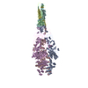
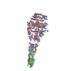
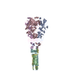
+ Open data
- Basic information
| Entry | Database: EMDB / ID: EMD-20698 | |||||||||
|---|---|---|---|---|---|---|---|---|---|---|
| Title | Structure of Francisella PdpA-VgrG Complex, half-lidded | |||||||||
 Map data Map data | Francisella PdpA-VgrG Complex, half-lidded | |||||||||
 Sample Sample |
| |||||||||
 Keywords Keywords | T6SS Central Spike / Complex / TRANSPORT PROTEIN | |||||||||
| Function / homology | symbiont cell surface / host cell cytoplasm / Uncharacterized protein / Uncharacterized protein Function and homology information Function and homology information | |||||||||
| Biological species |  Francisella tularensis subsp. novicida U112 (bacteria) / Francisella tularensis subsp. novicida U112 (bacteria) /  Francisella tularensis subsp. novicida (strain U112) (bacteria) Francisella tularensis subsp. novicida (strain U112) (bacteria) | |||||||||
| Method | single particle reconstruction / cryo EM / Resolution: 3.98 Å | |||||||||
 Authors Authors | Yang X / Clemens DL | |||||||||
| Funding support |  United States, 1 items United States, 1 items
| |||||||||
 Citation Citation |  Journal: Structure / Year: 2019 Journal: Structure / Year: 2019Title: Atomic Structure of the Francisella T6SS Central Spike Reveals a Unique α-Helical Lid and a Putative Cargo. Authors: Xue Yang / Daniel L Clemens / Bai-Yu Lee / Yanxiang Cui / Z Hong Zhou / Marcus A Horwitz /   Abstract: Francisella bacteria rely on a phylogenetically distinct type VI secretion system (T6SS) to escape host phagosomes and cause the fatal disease tularemia, but the structural and molecular mechanisms ...Francisella bacteria rely on a phylogenetically distinct type VI secretion system (T6SS) to escape host phagosomes and cause the fatal disease tularemia, but the structural and molecular mechanisms involved are unknown. Here we report the atomic structure of the Francisella T6SS central spike complex, obtained by cryo-electron microscopy. Our structural and functional studies demonstrate that, unlike the single-protein spike composition of other T6SS subtypes, Francisella T6SS's central spike is formed by two proteins, PdpA and VgrG, akin to T4-bacteriophage gp27 and gp5, respectively, and that PdpA has unique characteristics, including a putative cargo within its cavity and an N-terminal helical lid. Structure-guided mutagenesis demonstrates that the PdpA N-terminal lid and C-terminal spike are essential to Francisella T6SS function. PdpA is thus both an adaptor, connecting VgrG to the tube, and a likely carrier of secreted cargo. These findings are important to understanding Francisella pathogenicity and designing therapeutics to combat tularemia. | |||||||||
| History |
|
- Structure visualization
| Movie |
 Movie viewer Movie viewer |
|---|---|
| Structure viewer | EM map:  SurfView SurfView Molmil Molmil Jmol/JSmol Jmol/JSmol |
- Downloads & links
-EMDB archive
| Map data |  emd_20698.map.gz emd_20698.map.gz | 9.6 MB |  EMDB map data format EMDB map data format | |
|---|---|---|---|---|
| Header (meta data) |  emd-20698-v30.xml emd-20698-v30.xml emd-20698.xml emd-20698.xml | 12.7 KB 12.7 KB | Display Display |  EMDB header EMDB header |
| Images |  emd_20698.png emd_20698.png | 69.4 KB | ||
| Filedesc metadata |  emd-20698.cif.gz emd-20698.cif.gz | 5.7 KB | ||
| Archive directory |  http://ftp.pdbj.org/pub/emdb/structures/EMD-20698 http://ftp.pdbj.org/pub/emdb/structures/EMD-20698 ftp://ftp.pdbj.org/pub/emdb/structures/EMD-20698 ftp://ftp.pdbj.org/pub/emdb/structures/EMD-20698 | HTTPS FTP |
-Validation report
| Summary document |  emd_20698_validation.pdf.gz emd_20698_validation.pdf.gz | 350.6 KB | Display |  EMDB validaton report EMDB validaton report |
|---|---|---|---|---|
| Full document |  emd_20698_full_validation.pdf.gz emd_20698_full_validation.pdf.gz | 350.2 KB | Display | |
| Data in XML |  emd_20698_validation.xml.gz emd_20698_validation.xml.gz | 6.8 KB | Display | |
| Data in CIF |  emd_20698_validation.cif.gz emd_20698_validation.cif.gz | 7.7 KB | Display | |
| Arichive directory |  https://ftp.pdbj.org/pub/emdb/validation_reports/EMD-20698 https://ftp.pdbj.org/pub/emdb/validation_reports/EMD-20698 ftp://ftp.pdbj.org/pub/emdb/validation_reports/EMD-20698 ftp://ftp.pdbj.org/pub/emdb/validation_reports/EMD-20698 | HTTPS FTP |
-Related structure data
| Related structure data |  6u9gMC  6u9eC  6u9fC C: citing same article ( M: atomic model generated by this map |
|---|---|
| Similar structure data |
- Links
Links
| EMDB pages |  EMDB (EBI/PDBe) / EMDB (EBI/PDBe) /  EMDataResource EMDataResource |
|---|
- Map
Map
| File |  Download / File: emd_20698.map.gz / Format: CCP4 / Size: 163.6 MB / Type: IMAGE STORED AS FLOATING POINT NUMBER (4 BYTES) Download / File: emd_20698.map.gz / Format: CCP4 / Size: 163.6 MB / Type: IMAGE STORED AS FLOATING POINT NUMBER (4 BYTES) | ||||||||||||||||||||||||||||||||||||||||||||||||||||||||||||||||||||
|---|---|---|---|---|---|---|---|---|---|---|---|---|---|---|---|---|---|---|---|---|---|---|---|---|---|---|---|---|---|---|---|---|---|---|---|---|---|---|---|---|---|---|---|---|---|---|---|---|---|---|---|---|---|---|---|---|---|---|---|---|---|---|---|---|---|---|---|---|---|
| Annotation | Francisella PdpA-VgrG Complex, half-lidded | ||||||||||||||||||||||||||||||||||||||||||||||||||||||||||||||||||||
| Voxel size | X=Y=Z: 1.07 Å | ||||||||||||||||||||||||||||||||||||||||||||||||||||||||||||||||||||
| Density |
| ||||||||||||||||||||||||||||||||||||||||||||||||||||||||||||||||||||
| Symmetry | Space group: 1 | ||||||||||||||||||||||||||||||||||||||||||||||||||||||||||||||||||||
| Details | EMDB XML:
CCP4 map header:
| ||||||||||||||||||||||||||||||||||||||||||||||||||||||||||||||||||||
-Supplemental data
- Sample components
-Entire : PdpA-VgrG complex
| Entire | Name: PdpA-VgrG complex |
|---|---|
| Components |
|
-Supramolecule #1: PdpA-VgrG complex
| Supramolecule | Name: PdpA-VgrG complex / type: complex / ID: 1 / Parent: 0 / Macromolecule list: all |
|---|---|
| Source (natural) | Organism:  Francisella tularensis subsp. novicida U112 (bacteria) Francisella tularensis subsp. novicida U112 (bacteria) |
-Macromolecule #1: PdpA
| Macromolecule | Name: PdpA / type: protein_or_peptide / ID: 1 / Number of copies: 3 / Enantiomer: LEVO |
|---|---|
| Source (natural) | Organism:  Francisella tularensis subsp. novicida (strain U112) (bacteria) Francisella tularensis subsp. novicida (strain U112) (bacteria)Strain: U112 |
| Molecular weight | Theoretical: 95.469961 KDa |
| Recombinant expression | Organism:  |
| Sequence | String: MIAVKDITDL NIQDIISQLT SEVINGDTTP SSAKFACEIN SYIINYNLSN INLINTQLKN TKILYRKGLI SKLDYEKYKR YCIISRFKN NIDEFILYFS TNYKDSQSLK IAIKELQNSC SSSLILELPH DYIRKIDVLL TSIDSAIQRS SDLNKTIIKQ L NKLRSSLS ...String: MIAVKDITDL NIQDIISQLT SEVINGDTTP SSAKFACEIN SYIINYNLSN INLINTQLKN TKILYRKGLI SKLDYEKYKR YCIISRFKN NIDEFILYFS TNYKDSQSLK IAIKELQNSC SSSLILELPH DYIRKIDVLL TSIDSAIQRS SDLNKTIIKQ L NKLRSSLS RYIGYNNVLQ KQEITINIKP INKNFELEDI SFVSTRNKQY FKHNSLTLKN PHIEKLEVCE NIYGINGWLT FD LAYINNH KDFNFLLSPN QPILLDIQIN DSFNFYKKES KKDHHKRTTR FIAIGFNSNS IDIHENFEYS IYSYTKNVSS GVK KFKIQF HDPLKALWTK HKPSYIALNK SLDDIFKDNF FFDSLFSLDT NKSNNLKIRI PQAFISTVNR NFYDFFIQQL EQNK CYLKY FCDKKSGKVS YHVVDQVDND LQRNIVNSDE DLKDKLSPYD ISCFKKQILI SNKSNFYVKE KNICPDVTLN TQRKE DRKI SDTLVKPFSS ILKDNLQSVE YIQSNNDDKQ EIITTGFEIL LTSRNTLPFL DTEITLSKLD NDQNYLLGAT DIKSLY ISQ RKLLFKRSKY CSKQLYENLH NFHYKSDSES DVYEKIAFTK YPSLTHDNSI TYKIKDYSNL TPEYPKYKSF SNFYING RI TIGENVNNDS KKAYKFFKNH KPEESSIAEF QENGEKGTSA ILNSKADILY AIEIAKEMLS DKSSDKPIIY LPLKVNIN S ANNQFIPLRN DDIILIEIQS FTKGEIIELI SNSAISTKKA QQQLLQRQLL GSKENCEMAY TQTSDSETFS LTQVNEDCE NSFLINDKKG IFLRYKSKGN UniProtKB: Uncharacterized protein |
-Macromolecule #2: VgrG
| Macromolecule | Name: VgrG / type: protein_or_peptide / ID: 2 / Number of copies: 3 / Enantiomer: LEVO |
|---|---|
| Source (natural) | Organism:  Francisella tularensis subsp. novicida (strain U112) (bacteria) Francisella tularensis subsp. novicida (strain U112) (bacteria)Strain: U112 |
| Molecular weight | Theoretical: 20.539779 KDa |
| Recombinant expression | Organism:  |
| Sequence | String: MDYKDDDDKD YKDDDDKDYK DDDDKGSKAD HIFNLEEQGL LIDIKDDSKG CTTKLESSGK ITHNATESIE SSADKQIIEN VKDSKISIT EKEILLATKK SSIMLSEDKI VIKIGNSLII LDDSNISLES ATINIKSSAN INIQASQNID IKSLNNSIKA D VNLNAEGL DVNIKGSVTA SIKGSAATMV G UniProtKB: Uncharacterized protein |
-Experimental details
-Structure determination
| Method | cryo EM |
|---|---|
 Processing Processing | single particle reconstruction |
| Aggregation state | particle |
- Sample preparation
| Buffer | pH: 7.5 |
|---|---|
| Grid | Model: Quantifoil R2/1 / Material: COPPER / Mesh: 300 / Pretreatment - Type: PLASMA CLEANING / Pretreatment - Time: 60 sec. |
| Vitrification | Cryogen name: ETHANE / Chamber humidity: 100 % / Chamber temperature: 298 K / Instrument: FEI VITROBOT MARK IV |
- Electron microscopy
| Microscope | FEI TITAN KRIOS |
|---|---|
| Image recording | Film or detector model: GATAN K2 SUMMIT (4k x 4k) / Detector mode: SUPER-RESOLUTION / Average electron dose: 47.0 e/Å2 |
| Electron beam | Acceleration voltage: 300 kV / Electron source:  FIELD EMISSION GUN FIELD EMISSION GUN |
| Electron optics | Illumination mode: FLOOD BEAM / Imaging mode: BRIGHT FIELD |
| Sample stage | Specimen holder model: FEI TITAN KRIOS AUTOGRID HOLDER / Cooling holder cryogen: NITROGEN |
| Experimental equipment |  Model: Titan Krios / Image courtesy: FEI Company |
- Image processing
| Startup model | Type of model: NONE |
|---|---|
| Final reconstruction | Applied symmetry - Point group: C3 (3 fold cyclic) / Resolution.type: BY AUTHOR / Resolution: 3.98 Å / Resolution method: FSC 0.143 CUT-OFF / Number images used: 10343 |
| Initial angle assignment | Type: ANGULAR RECONSTITUTION |
| Final angle assignment | Type: ANGULAR RECONSTITUTION |
-Atomic model buiding 1
| Refinement | Space: REAL |
|---|---|
| Output model |  PDB-6u9g: |
 Movie
Movie Controller


