National Institutes of Health/National Institute Of Allergy and Infectious Diseases
NIAID R01 AI123351
United States
National Institutes of Health/National Institute of General Medical Sciences
NIGMS P30 GM110761
United States
Citation
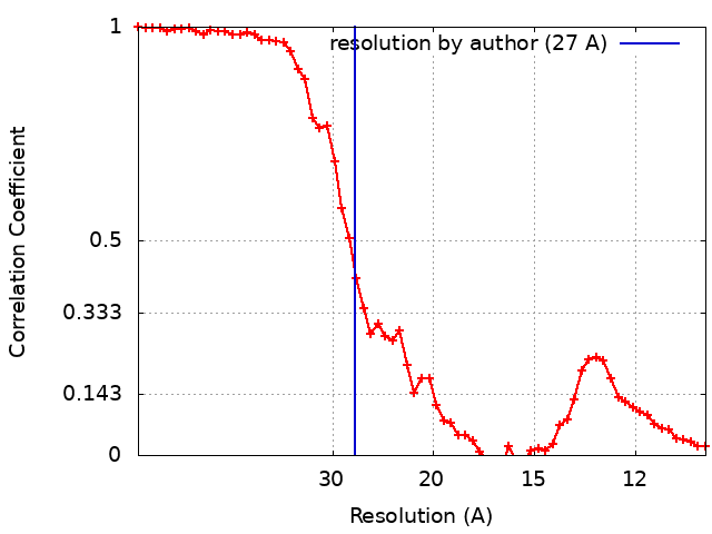
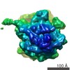
Journal: J Biol Chem / Year: 2019 Title: The cytoplasmic domain of MxiG interacts with MxiK and directs assembly of the sorting platform in the type III secretion system. Authors: Shoichi Tachiyama / Yunjie Chang / Meenakumari Muthuramalingam / Bo Hu / Michael L Barta / Wendy L Picking / Jun Liu / William D Picking / Abstract: Many Gram-negative bacteria use type III secretion systems (T3SSs) to inject virulence effector proteins into eukaryotic cells. The T3SS apparatus (T3SA) is structurally conserved among diverse ...Many Gram-negative bacteria use type III secretion systems (T3SSs) to inject virulence effector proteins into eukaryotic cells. The T3SS apparatus (T3SA) is structurally conserved among diverse bacterial pathogens and consists of a cytoplasmic sorting platform, an envelope-spanning basal body, and an extracellular needle with tip complex. The sorting platform is essential for effector recognition and powering secretion. Studies using bacterial "minicells" have revealed an unprecedented level of structural detail of the sorting platform; however, many of the structure-function relationships within this complex remain enigmatic. Here, we report on improved cryo-electron tomographic approaches to enhance the resolution of the T3SA sorting platform (at ≤2 nm resolution) done in concert with biochemical and genetic methods to define the sorting platform interactome and interactions with the T3SA inner membrane ring (IR). We observed that the sorting platform consists of "pods" with 6-fold symmetry that interact with the Spa47 ATPase via radial extensions comprising MxiN. Most importantly, MxiK maintained an interaction with the IR via specific interactions with the cytoplasmic domain of the IR protein MxiG (MxiG), which is a noncanonical forkhead-associated domain, and MxiK has an elongated structure that interacts with the IR via MxiG T4 lysozyme-mediated insertional mutagenesis of MxiK revealed its orientation within the sorting platform and enabled disruption of interactions with its binding partners, which abolished sorting platform assembly. Finally, a comparison with the homologous interactions in the T3SS sorting platform revealed clear differences in their IR-sorting platform interfaces that have possible mechanistic implications.
History
Deposition
Aug 15, 2019
-
Header (metadata) release
Sep 18, 2019
-
Map release
Nov 6, 2019
-
Update
Dec 25, 2019
-
Current status
Dec 25, 2019
Processing site: RCSB / Status: Released
-
Structure visualization
Movie
Surface view with section colored by density value
In the structure databanks used in Yorodumi, some data are registered as the other names, "COVID-19 virus" and "2019-nCoV". Here are the details of the virus and the list of structure data.
Jan 31, 2019. EMDB accession codes are about to change! (news from PDBe EMDB page)
EMDB accession codes are about to change! (news from PDBe EMDB page)
The allocation of 4 digits for EMDB accession codes will soon come to an end. Whilst these codes will remain in use, new EMDB accession codes will include an additional digit and will expand incrementally as the available range of codes is exhausted. The current 4-digit format prefixed with “EMD-” (i.e. EMD-XXXX) will advance to a 5-digit format (i.e. EMD-XXXXX), and so on. It is currently estimated that the 4-digit codes will be depleted around Spring 2019, at which point the 5-digit format will come into force.
The EM Navigator/Yorodumi systems omit the EMD- prefix.
Related info.:Q: What is EMD? / ID/Accession-code notation in Yorodumi/EM Navigator
Yorodumi is a browser for structure data from EMDB, PDB, SASBDB, etc.
This page is also the successor to EM Navigator detail page, and also detail information page/front-end page for Omokage search.
The word "yorodu" (or yorozu) is an old Japanese word meaning "ten thousand". "mi" (miru) is to see.
Related info.:EMDB / PDB / SASBDB / Comparison of 3 databanks / Yorodumi Search / Aug 31, 2016. New EM Navigator & Yorodumi / Yorodumi Papers / Jmol/JSmol / Function and homology information / Changes in new EM Navigator and Yorodumi
 Movie
Movie Controller

 Yorodumi
 Open data
 Basic information
 Map data
 Sample
 Shigella flexneri 5a str. M90T (bacteria)
 Authors
 United States, 2 items
  Citation
 Structure visualization
 Movie viewer
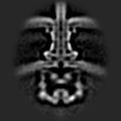
 Downloads & links
 emd_20611.png
 http://ftp.pdbj.org/pub/emdb/structures/EMD-20611
 Z (Sec.)
 Y (Row.)
 X (Col.)


 Sample components
 Processing
 Electron microscopy
 FIELD EMISSION GUN