National Institutes of Health/National Institute of General Medical Sciences
R01-GM112508
United States
National Institutes of Health/National Institute of General Medical Sciences
P50-GM082545
United States
National Institutes of Health/National Institute Of Allergy and Infectious Diseases
R01-AI150479
United States
National Institutes of Health/National Institute Of Allergy and Infectious Diseases
P50-AI150464
United States
Citation
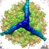
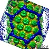
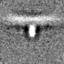
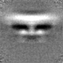
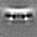
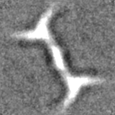
Journal: Sci Adv / Year: 2019 Title: Hierarchical assembly governs TRIM5α recognition of HIV-1 and retroviral capsids. Authors: Katarzyna A Skorupka / Marcin D Roganowicz / Devin E Christensen / Yueping Wan / Owen Pornillos / Barbie K Ganser-Pornillos / Abstract: TRIM5α is a restriction factor that senses incoming retrovirus cores through an unprecedented mechanism of nonself recognition. TRIM5α assembles a hexagonal lattice that avidly binds the capsid ...TRIM5α is a restriction factor that senses incoming retrovirus cores through an unprecedented mechanism of nonself recognition. TRIM5α assembles a hexagonal lattice that avidly binds the capsid shell, which surrounds and protects the virus core. The extent to which the TRIM lattice can cover the capsid and how TRIM5α directly contacts the capsid surface have not been established. Here, we apply cryo-electron tomography and subtomogram averaging to determine structures of TRIM5α bound to recombinant HIV-1 capsid assemblies. Our data support a mechanism of hierarchical assembly, in which a limited number of basal interaction modes are successively organized in increasingly higher-order structures that culminate in a TRIM5α cage surrounding a retroviral capsid. We further propose that cage formation explains the mechanism of restriction and provides the structural context that links capsid recognition to ubiquitin-dependent processes that disable the retrovirus.
History
Deposition
Aug 6, 2019
-
Header (metadata) release
Aug 28, 2019
-
Map release
Dec 18, 2019
-
Update
Dec 2, 2020
-
Current status
Dec 2, 2020
Processing site: RCSB / Status: Released
-
Structure visualization
Movie
Surface view with section colored by density value
In the structure databanks used in Yorodumi, some data are registered as the other names, "COVID-19 virus" and "2019-nCoV". Here are the details of the virus and the list of structure data.
Jan 31, 2019. EMDB accession codes are about to change! (news from PDBe EMDB page)
EMDB accession codes are about to change! (news from PDBe EMDB page)
The allocation of 4 digits for EMDB accession codes will soon come to an end. Whilst these codes will remain in use, new EMDB accession codes will include an additional digit and will expand incrementally as the available range of codes is exhausted. The current 4-digit format prefixed with “EMD-” (i.e. EMD-XXXX) will advance to a 5-digit format (i.e. EMD-XXXXX), and so on. It is currently estimated that the 4-digit codes will be depleted around Spring 2019, at which point the 5-digit format will come into force.
The EM Navigator/Yorodumi systems omit the EMD- prefix.
Related info.:Q: What is EMD? / ID/Accession-code notation in Yorodumi/EM Navigator
Yorodumi is a browser for structure data from EMDB, PDB, SASBDB, etc.
This page is also the successor to EM Navigator detail page, and also detail information page/front-end page for Omokage search.
The word "yorodu" (or yorozu) is an old Japanese word meaning "ten thousand". "mi" (miru) is to see.
Related info.:EMDB / PDB / SASBDB / Comparison of 3 databanks / Yorodumi Search / Aug 31, 2016. New EM Navigator & Yorodumi / Yorodumi Papers / Jmol/JSmol / Function and homology information / Changes in new EM Navigator and Yorodumi
 Movie
Movie Controller


 Open data
 Basic information
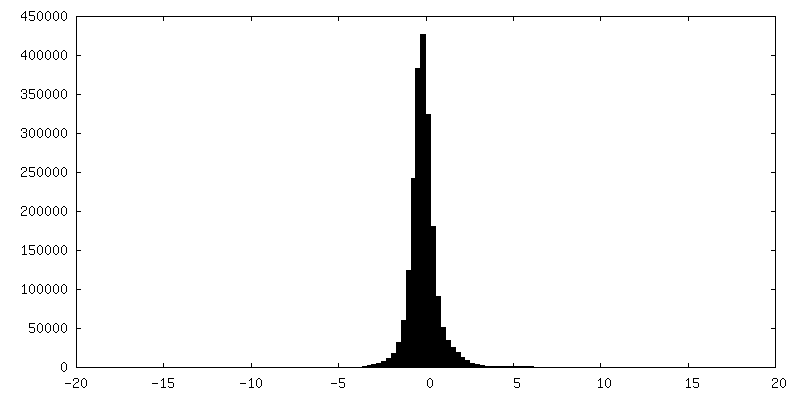
 Map data
 Sample
 Human immunodeficiency virus 1
 Authors
 United States, 4 items
  Citation
 Structure visualization
 Movie viewer
 Downloads & links
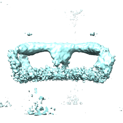 emd_20563.png
 http://ftp.pdbj.org/pub/emdb/structures/EMD-20563
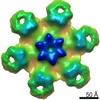

 Z (Sec.)
 Y (Row.)
 X (Col.)


 Sample components
 Processing
 Electron microscopy
 FIELD EMISSION GUN