National Institutes of Health/National Institute Of Allergy and Infectious Diseases
NIAID R01 AI123351
米国
National Institutes of Health/National Institute of General Medical Sciences
NIGMS P30 GM110761
米国
引用
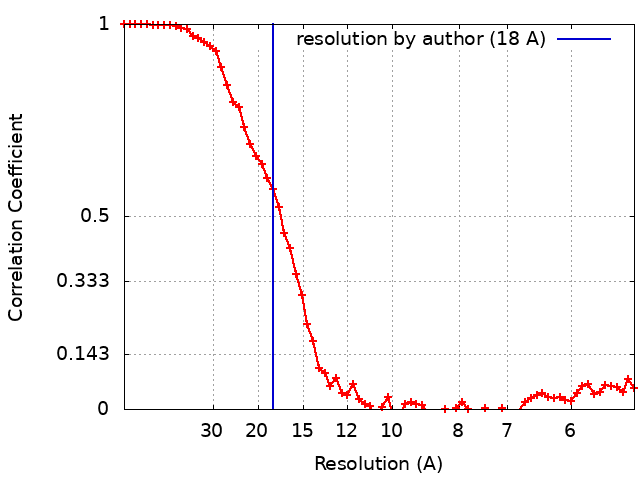
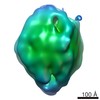
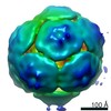
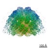
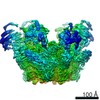
ジャーナル: J Biol Chem / 年: 2019 タイトル: The cytoplasmic domain of MxiG interacts with MxiK and directs assembly of the sorting platform in the type III secretion system. 著者: Shoichi Tachiyama / Yunjie Chang / Meenakumari Muthuramalingam / Bo Hu / Michael L Barta / Wendy L Picking / Jun Liu / William D Picking / 要旨: Many Gram-negative bacteria use type III secretion systems (T3SSs) to inject virulence effector proteins into eukaryotic cells. The T3SS apparatus (T3SA) is structurally conserved among diverse ...Many Gram-negative bacteria use type III secretion systems (T3SSs) to inject virulence effector proteins into eukaryotic cells. The T3SS apparatus (T3SA) is structurally conserved among diverse bacterial pathogens and consists of a cytoplasmic sorting platform, an envelope-spanning basal body, and an extracellular needle with tip complex. The sorting platform is essential for effector recognition and powering secretion. Studies using bacterial "minicells" have revealed an unprecedented level of structural detail of the sorting platform; however, many of the structure-function relationships within this complex remain enigmatic. Here, we report on improved cryo-electron tomographic approaches to enhance the resolution of the T3SA sorting platform (at ≤2 nm resolution) done in concert with biochemical and genetic methods to define the sorting platform interactome and interactions with the T3SA inner membrane ring (IR). We observed that the sorting platform consists of "pods" with 6-fold symmetry that interact with the Spa47 ATPase via radial extensions comprising MxiN. Most importantly, MxiK maintained an interaction with the IR via specific interactions with the cytoplasmic domain of the IR protein MxiG (MxiG), which is a noncanonical forkhead-associated domain, and MxiK has an elongated structure that interacts with the IR via MxiG T4 lysozyme-mediated insertional mutagenesis of MxiK revealed its orientation within the sorting platform and enabled disruption of interactions with its binding partners, which abolished sorting platform assembly. Finally, a comparison with the homologous interactions in the T3SS sorting platform revealed clear differences in their IR-sorting platform interfaces that have possible mechanistic implications.
 ムービー
ムービー コントローラー


 データを開く
 基本情報
基本情報 マップデータ
 試料
試料 Shigella flexneri (フレクスナー赤痢菌)
 データ登録者
 米国, 2件
  引用
引用 構造の表示
構造の表示 ムービービューア
 ダウンロードとリンク
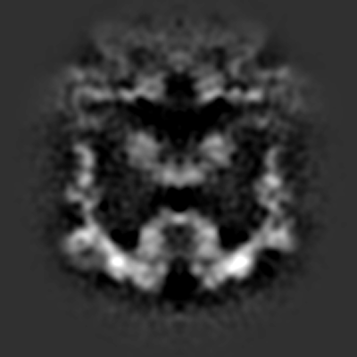 emd_20561.png
 http://ftp.pdbj.org/pub/emdb/structures/EMD-20561
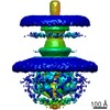
 Z (Sec.)
 Y (Row.)
 X (Col.)


 試料の構成要素
 解析
解析 電子顕微鏡法
 FIELD EMISSION GUN