 Movie
Movie Controller


+ Open data

- Basic information
| Entry | Database: EMDB / ID: EMD-20477 | |||||||||
|---|---|---|---|---|---|---|---|---|---|---|
| Title | Attachment of STIV to Sulfolobus pili | |||||||||
 Map data Map data | Attachment of Sulfolobus Turreted Icosahedral Virus (STIV) to Sulfolobus pili | |||||||||
 Sample Sample |
| |||||||||
| Biological species |    Sulfolobus turreted icosahedral virus 1 Sulfolobus turreted icosahedral virus 1 | |||||||||
| Method | subtomogram averaging / cryo EM / Resolution: 20.0 Å | |||||||||
 Authors Authors | Lawrence CM / Young MA / Hartman RA | |||||||||
| Funding support |  United States, 2 items United States, 2 items
| |||||||||
 Citation Citation | Journal: Structure / Year: 2019 Title: The Molecular Mechanism of Cellular Attachment for an Archaeal Virus. Authors: Ross Hartman / Brian J Eilers / Daniel Bollschweiler / Jacob H Munson-McGee / Harald Engelhardt / Mark J Young / C Martin Lawrence /  Abstract: Sulfolobus turreted icosahedral virus (STIV) is a model archaeal virus and member of the PRD1-adenovirus lineage. Although STIV employs pyramidal lysis structures to exit the host, knowledge of the ...Sulfolobus turreted icosahedral virus (STIV) is a model archaeal virus and member of the PRD1-adenovirus lineage. Although STIV employs pyramidal lysis structures to exit the host, knowledge of the viral entry process is lacking. We therefore initiated studies on STIV attachment and entry. Negative stain and cryoelectron micrographs showed virion attachment to pili-like structures emanating from the Sulfolobus host. Tomographic reconstruction and sub-tomogram averaging revealed pili recognition by the STIV C381 turret protein. Specifically, the triple jelly roll structure of C381 determined by X-ray crystallography shows that pilus recognition is mediated by conserved surface residues in the second and third domains. In addition, the STIV petal protein (C557), when present, occludes the pili binding site, suggesting that it functions as a maturation protein. Combined, these results demonstrate a role for the namesake STIV turrets in initial cellular attachment and provide the first molecular model for viral attachment in the archaeal domain of life. | |||||||||
| History |
|
- Structure visualization
| Movie |
 Movie viewer Movie viewer |
|---|---|
| Structure viewer | EM map: SurfViewMolmilJmol/JSmol |
| Supplemental images |

- Downloads & links
-EMDB archive
| Map data | emd_20477.map.gz | 20.3 MB | EMDB map data format | |
|---|---|---|---|---|
| Header (meta data) | emd-20477-v30.xmlemd-20477.xml | 10.1 KB 10.1 KB | Display Display | EMDB header |
| Images | 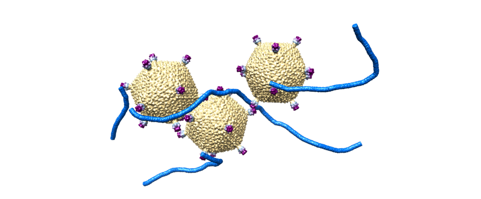 emd_20477.png emd_20477.png | 180.6 KB | ||
| Archive directory |  http://ftp.pdbj.org/pub/emdb/structures/EMD-20477ftp://ftp.pdbj.org/pub/emdb/structures/EMD-20477 http://ftp.pdbj.org/pub/emdb/structures/EMD-20477ftp://ftp.pdbj.org/pub/emdb/structures/EMD-20477 | HTTPS FTP |
-Links
| EMDB pages | EMDB (EBI/PDBe) / EMDataResource |
|---|
-Map
| File | Download / File: emd_20477.map.gz / Format: CCP4 / Size: 3.7 GB / Type: IMAGE STORED AS FLOATING POINT NUMBER (4 BYTES) | ||||||||||||||||||||||||||||||||||||||||||||||||||||||||||||||||||||
|---|---|---|---|---|---|---|---|---|---|---|---|---|---|---|---|---|---|---|---|---|---|---|---|---|---|---|---|---|---|---|---|---|---|---|---|---|---|---|---|---|---|---|---|---|---|---|---|---|---|---|---|---|---|---|---|---|---|---|---|---|---|---|---|---|---|---|---|---|---|
| Annotation | Attachment of Sulfolobus Turreted Icosahedral Virus (STIV) to Sulfolobus pili | ||||||||||||||||||||||||||||||||||||||||||||||||||||||||||||||||||||
| Projections & slices | Image control
Images are generated by Spider. generated in cubic-lattice coordinate | ||||||||||||||||||||||||||||||||||||||||||||||||||||||||||||||||||||
| Voxel size | X=Y=Z: 4.5 Å | ||||||||||||||||||||||||||||||||||||||||||||||||||||||||||||||||||||
| Density |
| ||||||||||||||||||||||||||||||||||||||||||||||||||||||||||||||||||||
| Symmetry | Space group: 1 | ||||||||||||||||||||||||||||||||||||||||||||||||||||||||||||||||||||
| Details | EMDB XML:
CCP4 map header:
| ||||||||||||||||||||||||||||||||||||||||||||||||||||||||||||||||||||
 Z (Sec.)
 Y (Row.)
 X (Col.)


-Supplemental data
- Sample components
-Entire : Sulfolobus turreted icosahedral virus 1
| Entire | Name: Sulfolobus turreted icosahedral virus 1 |
|---|---|
| Components |
|
-Supramolecule #1: Sulfolobus turreted icosahedral virus 1
| Supramolecule | Name: Sulfolobus turreted icosahedral virus 1 / type: virus / ID: 1 / Parent: 0 / NCBI-ID: 269145 / Sci species name: Sulfolobus turreted icosahedral virus 1 / Virus type: VIRION / Virus isolate: STRAIN / Virus enveloped: Yes / Virus empty: No |
|---|---|
| Host (natural) | Organism:  Saccharolobus solfataricus P2 (archaea) / Strain: P2^3 Saccharolobus solfataricus P2 (archaea) / Strain: P2^3 |
| Virus shell | Shell ID: 1 / Diameter: 740.0 Å / T number (triangulation number): 31 |
-Experimental details
-Structure determination
| Method | cryo EM |
|---|---|
 Processing Processing | subtomogram averaging |
| Aggregation state | 3D array |
-Sample preparation
| Buffer | pH: 2.5 / Component - Concentration: 5.0 mM / Component - Formula: C6H8O7 / Component - Name: Citric Acid |
|---|---|
| Grid | Model: Quantifoil R2/4 / Material: COPPER / Mesh: 200 |
| Vitrification | Cryogen name: ETHANE-PROPANE / Instrument: FEI VITROBOT MARK III |
| Details | 1 ml S. solfactaricus cells at approximately 5E10^8 cells/ml was combined with 100 ul of 1.88 mg/ml virus and incubated for 20 minutes at 80C. Unbound virus was removed by centrifugation. |
- Electron microscopy
| Microscope | FEI TITAN KRIOS |
|---|---|
| Specialist optics | Phase plate: VOLTA PHASE PLATE |
| Image recording | Film or detector model: GATAN K2 SUMMIT (4k x 4k) / Detector mode: COUNTING / Average electron dose: 60.0 e/Å2 |
| Electron beam | Acceleration voltage: 300 kV / Electron source:  FIELD EMISSION GUN FIELD EMISSION GUN |
| Electron optics | Illumination mode: FLOOD BEAM / Imaging mode: BRIGHT FIELD |
| Sample stage | Cooling holder cryogen: NITROGEN |
| Experimental equipment |  Model: Titan Krios / Image courtesy: FEI Company |
-Image processing
| Final reconstruction | Applied symmetry - Point group: I (icosahedral) / Resolution.type: BY AUTHOR / Resolution: 20.0 Å / Resolution method: FSC 0.143 CUT-OFF / Software - Name: PEET / Number subtomograms used: 7 |
|---|---|
| Extraction | Number tomograms: 1 / Number images used: 3 / Software - Name: PEET |
| Final angle assignment | Type: NOT APPLICABLE |
-Atomic model buiding 1
| Refinement | Protocol: RIGID BODY FIT |
|---|
