 Movie
Movie Controller


+ Open data

- Basic information
| Entry | Database: EMDB / ID: EMD-20024 | ||||||||||||||||||||||||
|---|---|---|---|---|---|---|---|---|---|---|---|---|---|---|---|---|---|---|---|---|---|---|---|---|---|
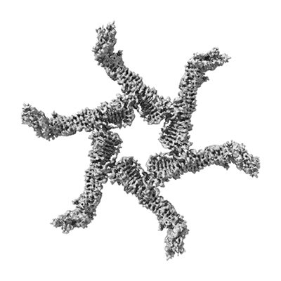
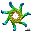
| Title | Cryo-EM structure of Helicobacter pylori VacA hexamer | ||||||||||||||||||||||||
 Map data Map data | Cryo-EM structure of Helicobacter pylori VacA hexamer | ||||||||||||||||||||||||
 Sample Sample |
| ||||||||||||||||||||||||
 Keywords Keywords | VacA / TOXIN | ||||||||||||||||||||||||
| Function / homology |  Function and homology information Function and homology informationcell outer membrane / toxin activity / periplasmic space / cell surface / extracellular region Similarity search - Function | ||||||||||||||||||||||||
| Biological species |   Helicobacter pylori (bacteria) Helicobacter pylori (bacteria) | ||||||||||||||||||||||||
| Method | single particle reconstruction / cryo EM / Resolution: 3.8 Å | ||||||||||||||||||||||||
 Authors Authors | Erwin AL / Cover TL | ||||||||||||||||||||||||
| Funding support |  United States, 7 items United States, 7 items
| ||||||||||||||||||||||||
 Citation Citation | Journal: J Mol Biol / Year: 2019 Title: Cryo-EM Analysis Reveals Structural Basis of Helicobacter pylori VacA Toxin Oligomerization. Authors: Min Su / Amanda L Erwin / Anne M Campbell / Tasia M Pyburn / Lauren E Salay / Jessica L Hanks / D Borden Lacy / David L Akey / Timothy L Cover / Melanie D Ohi / Abstract: Helicobacter pylori colonizes the human stomach and contributes to the development of gastric cancer and peptic ulcer disease. H. pylori secretes a pore-forming toxin called vacuolating cytotoxin A ...Helicobacter pylori colonizes the human stomach and contributes to the development of gastric cancer and peptic ulcer disease. H. pylori secretes a pore-forming toxin called vacuolating cytotoxin A (VacA), which contains two domains (p33 and p55) and assembles into oligomeric structures. Using single-particle cryo-electron microscopy, we have determined low-resolution structures of a VacA dodecamer and heptamer, as well as a 3.8-Å structure of the VacA hexamer. These analyses show that VacA p88 consists predominantly of a right-handed beta-helix that extends from the p55 domain into the p33 domain. We map the regions of p33 and p55 involved in hexamer assembly, model how interactions between protomers support heptamer formation, and identify surfaces of VacA that likely contact membrane. This work provides structural insights into the process of VacA oligomerization and identifies regions of VacA protomers that are predicted to contact the host cell surface during channel formation. | ||||||||||||||||||||||||
| History |
|
- Structure visualization
| Movie |
Movie viewer |
|---|---|
| Structure viewer | EM map: SurfViewMolmilJmol/JSmol |
| Supplemental images |
- Downloads & links
-EMDB archive
| Map data | emd_20024.map.gz | 115.9 MB | EMDB map data format | |
|---|---|---|---|---|
| Header (meta data) | emd-20024-v30.xmlemd-20024.xml | 12.4 KB 12.4 KB | Display Display | EMDB header |
| Images |  emd_20024.png emd_20024.png | 37.5 KB | ||
| Filedesc metadata | emd-20024.cif.gz | 5.1 KB | ||
| Archive directory |  http://ftp.pdbj.org/pub/emdb/structures/EMD-20024ftp://ftp.pdbj.org/pub/emdb/structures/EMD-20024 http://ftp.pdbj.org/pub/emdb/structures/EMD-20024ftp://ftp.pdbj.org/pub/emdb/structures/EMD-20024 | HTTPS FTP |
-Related structure data
| Related structure data | 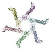 6odyMC M: atomic model generated by this map C: citing same article ( |
|---|---|
| Similar structure data |

-Links
| EMDB pages | EMDB (EBI/PDBe) / EMDataResource |
|---|
-Map
| File | Download / File: emd_20024.map.gz / Format: CCP4 / Size: 125 MB / Type: IMAGE STORED AS FLOATING POINT NUMBER (4 BYTES) | ||||||||||||||||||||||||||||||||||||||||||||||||||||||||||||
|---|---|---|---|---|---|---|---|---|---|---|---|---|---|---|---|---|---|---|---|---|---|---|---|---|---|---|---|---|---|---|---|---|---|---|---|---|---|---|---|---|---|---|---|---|---|---|---|---|---|---|---|---|---|---|---|---|---|---|---|---|---|
| Annotation | Cryo-EM structure of Helicobacter pylori VacA hexamer | ||||||||||||||||||||||||||||||||||||||||||||||||||||||||||||
| Projections & slices | Image control
Images are generated by Spider. | ||||||||||||||||||||||||||||||||||||||||||||||||||||||||||||
| Voxel size | X=Y=Z: 1.25 Å | ||||||||||||||||||||||||||||||||||||||||||||||||||||||||||||
| Density |
| ||||||||||||||||||||||||||||||||||||||||||||||||||||||||||||
| Symmetry | Space group: 1 | ||||||||||||||||||||||||||||||||||||||||||||||||||||||||||||
| Details | EMDB XML:
CCP4 map header:
| ||||||||||||||||||||||||||||||||||||||||||||||||||||||||||||
 Z (Sec.)
 Y (Row.)
 X (Col.)


-Supplemental data
- Sample components
-Entire : Vacuolating cytotoxin A
| Entire | Name: Vacuolating cytotoxin A |
|---|---|
| Components |
|
-Supramolecule #1: Vacuolating cytotoxin A
| Supramolecule | Name: Vacuolating cytotoxin A / type: complex / ID: 1 / Parent: 0 / Macromolecule list: all / Details: Hexamer |
|---|---|
| Source (natural) | Organism: Helicobacter pylori (bacteria) / Strain: s1/i1/m1 |
| Molecular weight | Theoretical: 528 KDa |
-Macromolecule #1: Vacuolating cytotoxin autotransporter
| Macromolecule | Name: Vacuolating cytotoxin autotransporter / type: protein_or_peptide / ID: 1 / Number of copies: 6 / Enantiomer: LEVO |
|---|---|
| Source (natural) | Organism: Helicobacter pylori (bacteria) / Strain: s1/i1/m1 |
| Molecular weight | Theoretical: 70.392578 KDa |
| Sequence | String: (UNK)(UNK)(UNK)(UNK)(UNK)(UNK)(UNK)(UNK)(UNK)(UNK) (UNK)(UNK)(UNK)(UNK)(UNK)(UNK) (UNK)(UNK)(UNK) (UNK)(UNK)(UNK)(UNK)(UNK)(UNK)(UNK)(UNK)(UNK)(UNK) (UNK)(UNK)(UNK) (UNK)(UNK)(UNK)(UNK)(UNK) ...String: (UNK)(UNK)(UNK)(UNK)(UNK)(UNK)(UNK)(UNK)(UNK)(UNK) (UNK)(UNK)(UNK)(UNK)(UNK)(UNK) (UNK)(UNK)(UNK) (UNK)(UNK)(UNK)(UNK)(UNK)(UNK)(UNK)(UNK)(UNK)(UNK) (UNK)(UNK)(UNK) (UNK)(UNK)(UNK)(UNK)(UNK)(UNK) (UNK)(UNK)(UNK)(UNK)(UNK)(UNK)(UNK)(UNK)(UNK)(UNK) (UNK)(UNK)(UNK)(UNK)(UNK)(UNK)(UNK)(UNK)(UNK) (UNK)(UNK)(UNK)(UNK)(UNK)(UNK)(UNK) (UNK)(UNK) (UNK)(UNK)(UNK)(UNK)(UNK)(UNK)(UNK)(UNK)(UNK)(UNK) (UNK)(UNK)(UNK)(UNK) (UNK)(UNK)(UNK)(UNK)(UNK) (UNK)(UNK)(UNK)(UNK)(UNK)(UNK)(UNK)(UNK)(UNK)(UNK) (UNK) (UNK)(UNK)(UNK)(UNK)(UNK)(UNK)(UNK)(UNK) (UNK)(UNK)(UNK)(UNK)(UNK)(UNK)(UNK)(UNK) (UNK) (UNK)(UNK)(UNK)(UNK)(UNK)(UNK)(UNK)(UNK)(UNK)(UNK) (UNK)(UNK)(UNK)(UNK)(UNK) (UNK)(UNK)(UNK)(UNK) (UNK)(UNK)(UNK)(UNK)(UNK)(UNK)(UNK)(UNK)(UNK)(UNK) (UNK)(UNK) (UNK)(UNK)(UNK)(UNK)(UNK)(UNK)(UNK) (UNK)(UNK)(UNK)(UNK)(UNK)(UNK)(UNK)(UNK)(UNK) (UNK)(UNK)(UNK)(UNK)(UNK)(UNK)(UNK)(UNK)(UNK)(UNK) (UNK)(UNK)(UNK)(UNK)(UNK)(UNK) (UNK)(UNK)(UNK) (UNK)(UNK)(UNK)(UNK)(UNK)(UNK)(UNK)(UNK)(UNK)(UNK) (UNK)(UNK)(UNK) (UNK)(UNK)(UNK)(UNK)(UNK)(UNK) (UNK)(UNK)(UNK)(UNK)(UNK)(UNK)(UNK)(UNK)(UNK)(UNK) (UNK)(UNK)(UNK)(UNK)(UNK)(UNK)(UNK)(UNK)(UNK) (UNK)(UNK)(UNK)(UNK)(UNK)(UNK)(UNK) (UNK)(UNK) (UNK)(UNK)(UNK)QPTQVID GPFAGGKDTV VNIDRINTKA DGTIKVGGFK ASLTTNAAHL NIGKGGVN L SNQASGRTLL VENLTGNITV DGPLRVNNQV GGYALAGSSA NFEFKAGVDT KNGTATFNND ISLGRFVNLK VDAHTANFK GIDTGNGGFN TLDFSGVTNK VNINKLITAS TNVAVKNFNI NELIVKTNGV SVGEYTHFSE DIGSQSRINT VRLETGTRSI FSGGVKFKS GEKLVIDEFY YSPWNYFDAR NIKNVEITRK FASSTPENPW GTSKLMFNNL TLGQNAVMDY SQFSNLTIQG D FINNQGTI NYLVRGGKVA TLNVGNAAAM MFNNDIDSAT GFYKPLIKIN SAQDLIKNTE HVLLKAKIIG YGNVSTGTNG IS NVNLEEQ FKERLALYNN NNRMDTCVVR NTDDIKACGM AIGNQSMVNN PDNYKYLIGK AWKNIGISKT ANGSKISVYY LGN STPTEN GGNTTNLPTN T UniProtKB: Vacuolating cytotoxin autotransporter |
-Experimental details
-Structure determination
| Method | cryo EM |
|---|---|
 Processing Processing | single particle reconstruction |
| Aggregation state | particle |
-Sample preparation
| Buffer | pH: 8 |
|---|---|
| Grid | Details: unspecified |
| Vitrification | Cryogen name: ETHANE |
- Electron microscopy
| Microscope | FEI TITAN KRIOS |
|---|---|
| Image recording | Film or detector model: GATAN K2 SUMMIT (4k x 4k) / Average electron dose: 48.0 e/Å2 |
| Electron beam | Acceleration voltage: 300 kV / Electron source:  FIELD EMISSION GUN FIELD EMISSION GUN |
| Electron optics | Illumination mode: FLOOD BEAM / Imaging mode: BRIGHT FIELD |
| Experimental equipment |  Model: Titan Krios / Image courtesy: FEI Company |
-Image processing
| Startup model | Type of model: OTHER / Details: Ab initio model |
|---|---|
| Final reconstruction | Resolution.type: BY AUTHOR / Resolution: 3.8 Å / Resolution method: FSC 0.143 CUT-OFF / Number images used: 133827 |
| Initial angle assignment | Type: MAXIMUM LIKELIHOOD |
| Final angle assignment | Type: PROJECTION MATCHING |
