 Movie
Movie Controller


+ Open data

- Basic information
| Entry |  | |||||||||
|---|---|---|---|---|---|---|---|---|---|---|
| Title | Plasmodium falciparum sporozoite actin determined in situ | |||||||||
 Map data Map data | ||||||||||
 Sample Sample |
| |||||||||
 Keywords Keywords | F-actin / actin / glideosome / STRUCTURAL PROTEIN | |||||||||
| Biological species |  | |||||||||
| Method | subtomogram averaging / cryo EM / Resolution: 28.0 Å | |||||||||
 Authors Authors | Prazak V / Ferreira JL | |||||||||
| Funding support |  France, France,  United Kingdom, 2 items United Kingdom, 2 items
| |||||||||
 Citation Citation | Journal: EMBO Rep / Year: 2025 Title: Molecular architecture of glideosome and nuclear F-actin in Plasmodium falciparum. Authors: Vojtěch Pražák / Daven Vasishtan / Kay Grünewald / Ross G Douglas / Josie L Ferreira /  Abstract: Actin-based motility is required for the transmission of malaria sporozoites. While this has been shown biochemically, filamentous actin has remained elusive and has not been directly visualised ...Actin-based motility is required for the transmission of malaria sporozoites. While this has been shown biochemically, filamentous actin has remained elusive and has not been directly visualised inside the parasite. Using focused ion beam milling and electron cryo-tomography, we studied dynamic actin filaments in unperturbed Plasmodium falciparum cells for the first time. This allowed us to dissect the assembly, path and fate of actin filaments during parasite gliding and determine a complete 3D model of F-actin within sporozoites. We observe micrometre long actin filaments, much longer than expected from in vitro studies. After their assembly at the parasite's apical end, actin filaments continue to grow as they are transported down the cell as part of the glideosome machinery, and are disassembled at the basal end in a rate-limiting step. Large pores in the IMC, constrained to the basal end, may facilitate actin exchange between the pellicular space and cytosol for recycling and maintenance of directional flow. The data also reveal striking actin bundles in the nucleus. Implications for motility and transmission are discussed. | |||||||||
| History |
|
- Structure visualization
| Supplemental images |
|---|

- Downloads & links
-EMDB archive
| Map data | emd_19898.map.gz | 224.5 KB |  EMDB map data format EMDB map data format | |
|---|---|---|---|---|
| Header (meta data) | emd-19898-v30.xmlemd-19898.xml | 15.1 KB 15.1 KB | Display Display | EMDB header |
| FSC (resolution estimation) | emd_19898_fsc.xml | 4.4 KB | Display | FSC data file |
| Images |  emd_19898.png emd_19898.png | 54.8 KB | ||
| Filedesc metadata | emd-19898.cif.gz | 4.3 KB | ||
| Others | emd_19898_additional_1.map.gzemd_19898_half_map_1.map.gzemd_19898_half_map_2.map.gz | 221.7 KB 222 KB 221.7 KB | ||
| Archive directory |  http://ftp.pdbj.org/pub/emdb/structures/EMD-19898ftp://ftp.pdbj.org/pub/emdb/structures/EMD-19898 http://ftp.pdbj.org/pub/emdb/structures/EMD-19898ftp://ftp.pdbj.org/pub/emdb/structures/EMD-19898 | HTTPS FTP |
-Links
| EMDB pages | EMDB (EBI/PDBe) / EMDataResource |
|---|
-Map
| File | Download / File: emd_19898.map.gz / Format: CCP4 / Size: 251 KB / Type: IMAGE STORED AS FLOATING POINT NUMBER (4 BYTES) | ||||||||||||||||||||||||||||||||||||
|---|---|---|---|---|---|---|---|---|---|---|---|---|---|---|---|---|---|---|---|---|---|---|---|---|---|---|---|---|---|---|---|---|---|---|---|---|---|
| Projections & slices | Image control
Images are generated by Spider. | ||||||||||||||||||||||||||||||||||||
| Voxel size | X=Y=Z: 6.672 Å | ||||||||||||||||||||||||||||||||||||
| Density |
| ||||||||||||||||||||||||||||||||||||
| Symmetry | Space group: 1 | ||||||||||||||||||||||||||||||||||||
| Details | EMDB XML:
|
 Z (Sec.)
 Y (Row.)
 X (Col.)


-Supplemental data
-Additional map: Unfiltered map
| File | emd_19898_additional_1.map | ||||||||||||
|---|---|---|---|---|---|---|---|---|---|---|---|---|---|
| Annotation | Unfiltered map | ||||||||||||
| Projections & Slices |
| ||||||||||||
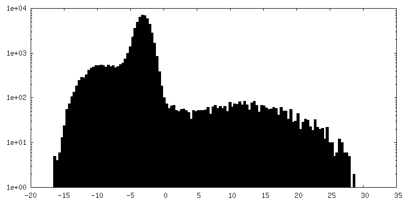
| Density Histograms |


-Half map: half map 2
| File | emd_19898_half_map_1.map | ||||||||||||
|---|---|---|---|---|---|---|---|---|---|---|---|---|---|
| Annotation | half map 2 | ||||||||||||
| Projections & Slices |
| ||||||||||||
| Density Histograms |


-Half map: half map 1
| File | emd_19898_half_map_2.map | ||||||||||||
|---|---|---|---|---|---|---|---|---|---|---|---|---|---|
| Annotation | half map 1 | ||||||||||||
| Projections & Slices |
| ||||||||||||
| Density Histograms |


- Sample components
-Entire : F-actin
| Entire | Name: F-actin |
|---|---|
| Components |
|
-Supramolecule #1: F-actin
| Supramolecule | Name: F-actin / type: organelle_or_cellular_component / ID: 1 / Parent: 0 |
|---|---|
| Source (natural) | Organism: |
-Experimental details
-Structure determination
| Method | cryo EM |
|---|---|
 Processing Processing | subtomogram averaging |
| Aggregation state | filament |
-Sample preparation
| Buffer | pH: 7.5 |
|---|---|
| Grid | Model: UltrAuFoil R1.2/1.3 / Material: GOLD / Mesh: 300 / Pretreatment - Type: GLOW DISCHARGE / Pretreatment - Time: 60 sec. |
| Vitrification | Cryogen name: ETHANE-PROPANE / Instrument: HOMEMADE PLUNGER |
| Details | Sporozoite stage parasites dissected from infected mosquito salivary glands were vitried on EM grids and FIB-milled. Actin was manually picked from celluar tomograms. |
- Electron microscopy
| Microscope | FEI TITAN KRIOS |
|---|---|
| Image recording | Film or detector model: GATAN K3 BIOQUANTUM (6k x 4k) / Average electron dose: 3.0 e/Å2 |
| Electron beam | Acceleration voltage: 300 kV / Electron source:  FIELD EMISSION GUN FIELD EMISSION GUN |
| Electron optics | C2 aperture diameter: 100.0 µm / Illumination mode: FLOOD BEAM / Imaging mode: BRIGHT FIELD / Nominal defocus max: 6.0 µm / Nominal defocus min: 3.0 µm |
| Experimental equipment |  Model: Titan Krios / Image courtesy: FEI Company |

