 Movie
Movie Controller


+ Open data

- Basic information
| Entry | 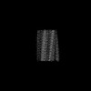 | |||||||||
|---|---|---|---|---|---|---|---|---|---|---|
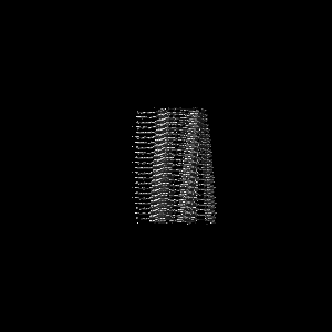
| Title | AL amyloid fibril from the FOR103 light chain | |||||||||
 Map data Map data | ||||||||||
 Sample Sample |
| |||||||||
 Keywords Keywords | Amyloid fibril / AL amyloid / PROTEIN FIBRIL | |||||||||
| Biological species |  Homo sapiens (human) Homo sapiens (human) | |||||||||
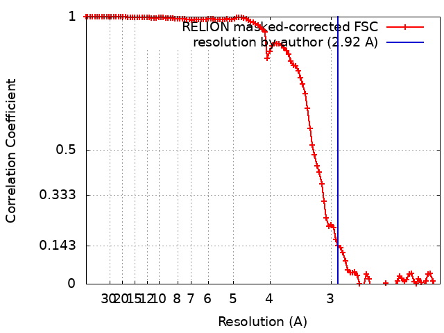
| Method | helical reconstruction / cryo EM / Resolution: 2.92 Å | |||||||||
 Authors Authors | Pfeiffer PB / Karimi-Farsijani S / Kupfer N / Schmidt M / Faendrich M | |||||||||
| Funding support |  Germany, 1 items Germany, 1 items
| |||||||||
 Citation Citation | Journal: Nat Commun / Year: 2024 Title: Light chain mutations contribute to defining the fibril morphology in systemic AL amyloidosis. Authors: Sara Karimi-Farsijani / Peter Benedikt Pfeiffer / Sambhasan Banerjee / Julian Baur / Lukas Kuhn / Niklas Kupfer / Ute Hegenbart / Stefan O Schönland / Sebastian Wiese / Christian Haupt / ...Authors: Sara Karimi-Farsijani / Peter Benedikt Pfeiffer / Sambhasan Banerjee / Julian Baur / Lukas Kuhn / Niklas Kupfer / Ute Hegenbart / Stefan O Schönland / Sebastian Wiese / Christian Haupt / Matthias Schmidt / Marcus Fändrich / Abstract: Systemic AL amyloidosis is one of the most frequently diagnosed forms of systemic amyloidosis. It arises from mutational changes in immunoglobulin light chains. To explore whether these mutations may ...Systemic AL amyloidosis is one of the most frequently diagnosed forms of systemic amyloidosis. It arises from mutational changes in immunoglobulin light chains. To explore whether these mutations may affect the structure of the formed fibrils, we determine and compare the fibril structures from several patients with cardiac AL amyloidosis. All patients are affected by light chains that contain an IGLV3-19 gene segment, and the deposited fibrils differ by the mutations within this common germ line background. Using cryo-electron microscopy, we here find different fibril structures in each patient. These data establish that the mutations of amyloidogenic light chains contribute to defining the fibril architecture and hence the structure of the pathogenic agent. | |||||||||
| History |
|
- Structure visualization
| Supplemental images |
|---|

- Downloads & links
-EMDB archive
| Map data | emd_19818.map.gz | 7.8 MB |  EMDB map data format EMDB map data format | |
|---|---|---|---|---|
| Header (meta data) | emd-19818-v30.xmlemd-19818.xml | 15.7 KB 15.7 KB | Display Display | EMDB header |
| FSC (resolution estimation) | emd_19818_fsc.xml | 10.6 KB | Display | FSC data file |
| Images | 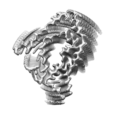 emd_19818.png emd_19818.png | 96 KB | ||
| Masks | emd_19818_msk_1.map | 103 MB | Mask map | |
| Filedesc metadata | emd-19818.cif.gz | 5.3 KB | ||
| Others | emd_19818_half_map_1.map.gzemd_19818_half_map_2.map.gz | 80.3 MB 80.3 MB | ||
| Archive directory |  http://ftp.pdbj.org/pub/emdb/structures/EMD-19818ftp://ftp.pdbj.org/pub/emdb/structures/EMD-19818 http://ftp.pdbj.org/pub/emdb/structures/EMD-19818ftp://ftp.pdbj.org/pub/emdb/structures/EMD-19818 | HTTPS FTP |
-Related structure data

-Links
| EMDB pages | EMDB (EBI/PDBe) / EMDataResource |
|---|
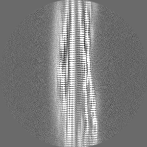
-Map
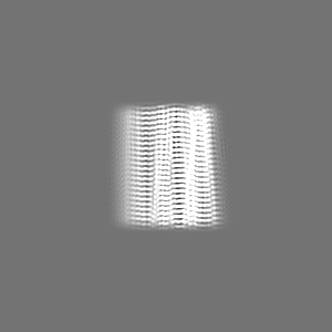
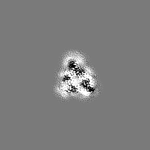
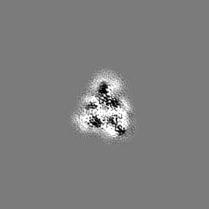
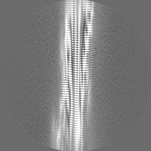
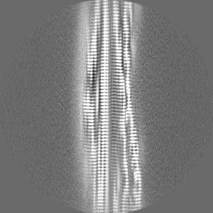
| File | Download / File: emd_19818.map.gz / Format: CCP4 / Size: 103 MB / Type: IMAGE STORED AS FLOATING POINT NUMBER (4 BYTES) | ||||||||||||||||||||||||||||||||||||
|---|---|---|---|---|---|---|---|---|---|---|---|---|---|---|---|---|---|---|---|---|---|---|---|---|---|---|---|---|---|---|---|---|---|---|---|---|---|
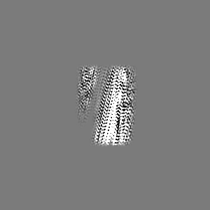
| Projections & slices | Image control
Images are generated by Spider. | ||||||||||||||||||||||||||||||||||||
| Voxel size | X=Y=Z: 1.04 Å | ||||||||||||||||||||||||||||||||||||
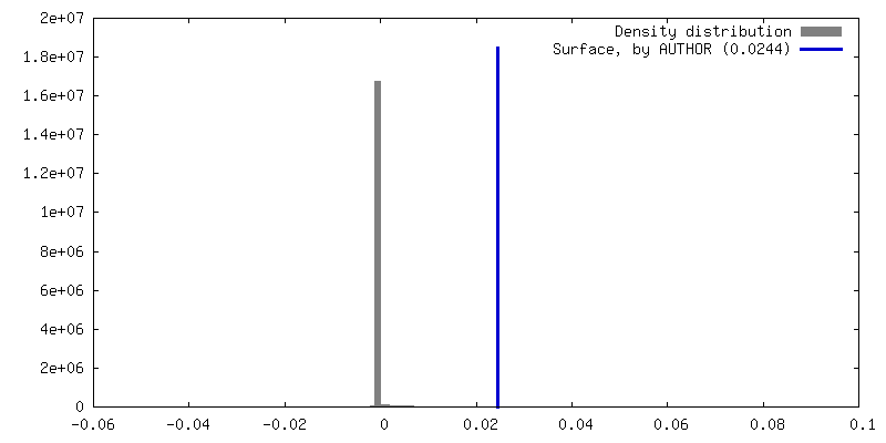
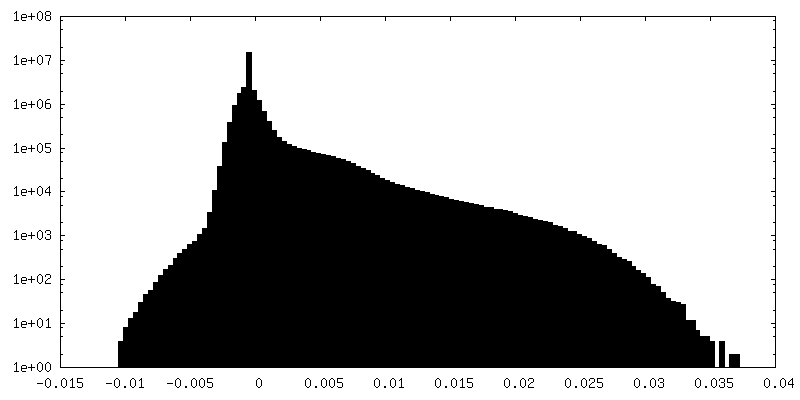
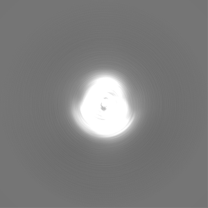
| Density |
| ||||||||||||||||||||||||||||||||||||
| Symmetry | Space group: 1 | ||||||||||||||||||||||||||||||||||||
| Details | EMDB XML:
|
 Z (Sec.)
 Y (Row.)
 X (Col.)


-Supplemental data
-Mask #1
| File | emd_19818_msk_1.map | ||||||||||||
|---|---|---|---|---|---|---|---|---|---|---|---|---|---|
| Projections & Slices |
| ||||||||||||
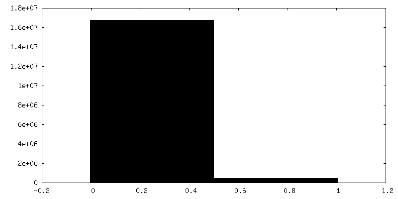
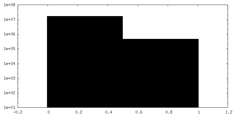
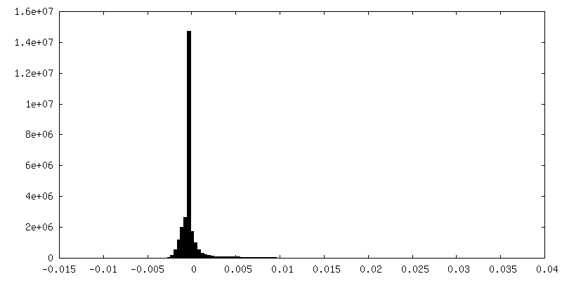
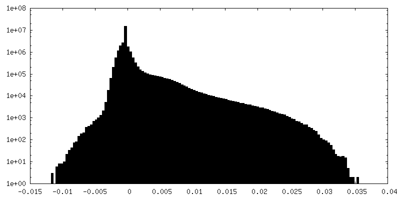
| Density Histograms |


-Half map: #1
| File | emd_19818_half_map_1.map | ||||||||||||
|---|---|---|---|---|---|---|---|---|---|---|---|---|---|
| Projections & Slices |
| ||||||||||||
| Density Histograms |

-Half map: #2
| File | emd_19818_half_map_2.map | ||||||||||||
|---|---|---|---|---|---|---|---|---|---|---|---|---|---|
| Projections & Slices |
| ||||||||||||
| Density Histograms |
- Sample components
-Entire : lambda 3 immunoglobulin light chain fragment
| Entire | Name: lambda 3 immunoglobulin light chain fragment |
|---|---|
| Components |
|
-Supramolecule #1: lambda 3 immunoglobulin light chain fragment
| Supramolecule | Name: lambda 3 immunoglobulin light chain fragment / type: complex / ID: 1 / Parent: 0 / Macromolecule list: all / Details: residues 2-116 |
|---|---|
| Source (natural) | Organism: Homo sapiens (human) |
-Macromolecule #1: lambda 3 immunoglobulin light chain fragment, residues 2-116
| Macromolecule | Name: lambda 3 immunoglobulin light chain fragment, residues 2-116 type: protein_or_peptide / ID: 1 / Number of copies: 6 / Enantiomer: LEVO |
|---|---|
| Source (natural) | Organism: Homo sapiens (human) |
| Molecular weight | Theoretical: 12.202262 KDa |
| Sequence | String: SELTQDPAVS VALGQTVRIT CQGDSLRSYY ASWYQQKSGQ APVLVIYSYN NRPSGIPDRF SGSNSGNTAS LTITGAQAED EADYYCNSR DSSGHHLVFG GGTKLTVLGQ PKAAPS |
-Experimental details
-Structure determination
| Method | cryo EM |
|---|---|
 Processing Processing | helical reconstruction |
| Aggregation state | helical array |
-Sample preparation
| Buffer | pH: 7 / Details: water |
|---|---|
| Grid | Model: C-flat-1.2/1.3 / Material: COPPER / Mesh: 400 / Support film - Material: CARBON / Support film - topology: HOLEY / Pretreatment - Type: GLOW DISCHARGE / Pretreatment - Time: 40 sec. |
| Vitrification | Cryogen name: ETHANE / Chamber humidity: 95 % / Instrument: LEICA PLUNGER |
- Electron microscopy
| Microscope | FEI TITAN KRIOS |
|---|---|
| Image recording | Film or detector model: GATAN K2 QUANTUM (4k x 4k) / Detector mode: COUNTING / Number grids imaged: 1 / Number real images: 2788 / Average exposure time: 10.0 sec. / Average electron dose: 53.7 e/Å2 |
| Electron beam | Acceleration voltage: 300 kV / Electron source:  FIELD EMISSION GUN FIELD EMISSION GUN |
| Electron optics | Illumination mode: FLOOD BEAM / Imaging mode: BRIGHT FIELD / Cs: 2.7 mm / Nominal defocus max: 2.5 µm / Nominal defocus min: 1.2 µm / Nominal magnification: 130000 |
| Sample stage | Cooling holder cryogen: NITROGEN |
| Experimental equipment |  Model: Titan Krios / Image courtesy: FEI Company |
+Image processing
-Atomic model buiding 1
| Refinement | Space: REAL / Protocol: BACKBONE TRACE / Target criteria: correlation coefficient |
|---|---|
| Output model |  PDB-9eme: |
