 Movie
Movie Controller


+ Open data

- Basic information
| Entry | 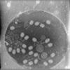 | |||||||||||||||
|---|---|---|---|---|---|---|---|---|---|---|---|---|---|---|---|---|
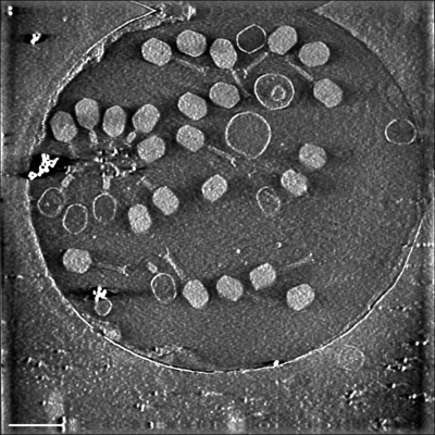
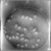
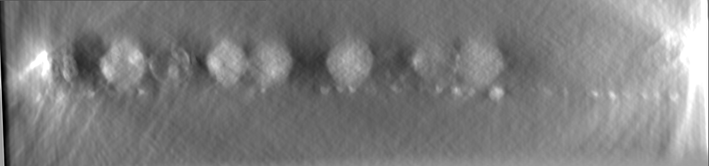
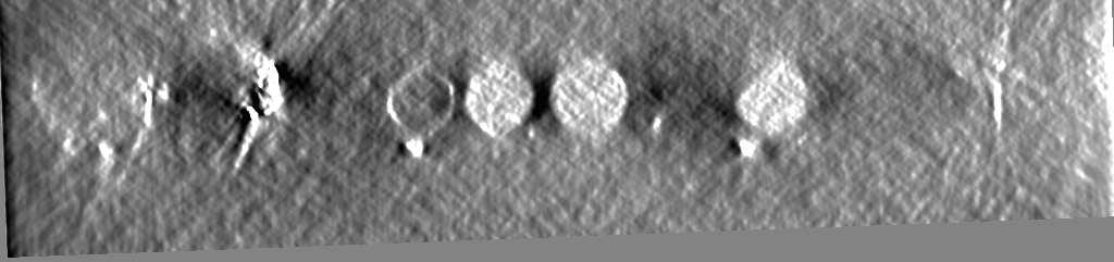
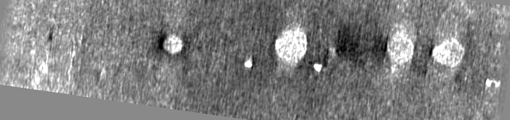
| Title | 4D_STEM cryo tomogram of t4 Phages produced by PCA2 | |||||||||||||||
 Map data Map data | 4D_STEM cryo tomogram of t4 Phages produced by PCA2 | |||||||||||||||
 Sample Sample |
| |||||||||||||||
 Keywords Keywords | 4D-STEM cryo tomography / Cryo-ET / CSTET / T4-Phage / iDPC / UNKNOWN FUNCTION | |||||||||||||||
| Biological species |  Escherichia phage T4 (virus) Escherichia phage T4 (virus) | |||||||||||||||
| Method | electron tomography / cryo EM | |||||||||||||||
 Authors Authors | Seifer S / Kirchweger P / Edel KM / Elbaum M | |||||||||||||||
| Funding support |  Austria, Austria,  Israel, European Union, 4 items Israel, European Union, 4 items
| |||||||||||||||
 Citation Citation | Journal: Microsc Microanal / Year: 2024 Title: Optimizing Contrast in Automated 4D STEM Cryotomography. Authors: Shahar Seifer / Peter Kirchweger / Karlina Maria Edel / Michael Elbaum / Abstract: 4D STEM is an emerging approach to electron microscopy. While it was developed principally for high-resolution studies in materials science, the possibility to collect the entire transmitted flux ...4D STEM is an emerging approach to electron microscopy. While it was developed principally for high-resolution studies in materials science, the possibility to collect the entire transmitted flux makes it attractive for cryomicroscopy in application to life science and radiation-sensitive materials where dose efficiency is of utmost importance. We present a workflow to acquire tomographic tilt series of 4D STEM data sets using a segmented diode and an ultrafast pixelated detector, demonstrating the methods using a specimen of a T4 bacteriophage. Full integration with the SerialEM platform conveniently provides all the tools for grid navigation and automation of the data collection. Scripts are provided to convert the raw data to mrc format files and further to generate a variety of modes representing both scattering and phase contrasts, including incoherent and annular bright field, integrated center of mass, and parallax decomposition of a simulated integrated differential phase contrast. Principal component analysis of virtual annular detectors proves particularly useful, and axial contrast is improved by 3D deconvolution with an optimized point spread function. Contrast optimization enables visualization of irregular features such as DNA strands and thin filaments of the phage tails, which would be lost upon averaging or imposition of an inappropriate symmetry. | |||||||||||||||
| History |
|
- Structure visualization
| Supplemental images |
|---|
- Downloads & links
-EMDB archive
| Map data | emd_19762.map.gz | 798.1 MB |  EMDB map data format EMDB map data format | |
|---|---|---|---|---|
| Header (meta data) | emd-19762-v30.xmlemd-19762.xml | 12.2 KB 12.2 KB | Display Display | EMDB header |
| Images |  emd_19762.png emd_19762.png | 188.4 KB | ||
| Filedesc metadata | emd-19762.cif.gz | 4.4 KB | ||
| Archive directory |  http://ftp.pdbj.org/pub/emdb/structures/EMD-19762ftp://ftp.pdbj.org/pub/emdb/structures/EMD-19762 http://ftp.pdbj.org/pub/emdb/structures/EMD-19762ftp://ftp.pdbj.org/pub/emdb/structures/EMD-19762 | HTTPS FTP |
-Related structure data
-Links
| EMDB pages | EMDB (EBI/PDBe) / EMDataResource |
|---|
-Map
| File | Download / File: emd_19762.map.gz / Format: CCP4 / Size: 964 MB / Type: IMAGE STORED AS FLOATING POINT NUMBER (4 BYTES) | ||||||||||||||||||||||||||||||||
|---|---|---|---|---|---|---|---|---|---|---|---|---|---|---|---|---|---|---|---|---|---|---|---|---|---|---|---|---|---|---|---|---|---|
| Annotation | 4D_STEM cryo tomogram of t4 Phages produced by PCA2 | ||||||||||||||||||||||||||||||||
| Projections & slices | Image control
Images are generated by Spider. generated in cubic-lattice coordinate | ||||||||||||||||||||||||||||||||
| Voxel size | X=Y=Z: 15.58 Å | ||||||||||||||||||||||||||||||||
| Density |
| ||||||||||||||||||||||||||||||||
| Symmetry | Space group: 1 | ||||||||||||||||||||||||||||||||
| Details | EMDB XML:
|
 Z (Sec.)
 Y (Row.)
 X (Col.)


-Supplemental data
- Sample components
-Entire : Escherichia phage T4
| Entire | Name: Escherichia phage T4 (virus) |
|---|---|
| Components |
|
-Supramolecule #1: Escherichia phage T4
| Supramolecule | Name: Escherichia phage T4 / type: virus / ID: 1 / Parent: 0 / Details: Isolated T4 Bacteriophage / NCBI-ID: 2681598 / Sci species name: Escherichia phage T4 / Virus type: VIROID / Virus isolate: STRAIN / Virus enveloped: Yes / Virus empty: No |
|---|---|
| Host (natural) | Organism:  |
-Supramolecule #2: Escherichia phage T4
| Supramolecule | Name: Escherichia phage T4 / type: organelle_or_cellular_component / ID: 2 / Parent: 1 |
|---|---|
| Source (natural) | Organism: Escherichia phage T4 (virus) |
-Experimental details
-Structure determination
| Method | cryo EM |
|---|---|
 Processing Processing | electron tomography |
| Aggregation state | particle |
-Sample preparation
| Buffer | pH: 7.4 / Details: 50 mM Tris pH 8 with 1 mM MgCl2 |
|---|---|
| Grid | Model: Quantifoil R1.2/1.3 / Material: COPPER / Mesh: 200 / Support film - #0 - Film type ID: 1 / Support film - #0 - Material: CARBON / Support film - #0 - topology: HOLEY / Support film - #1 - Film type ID: 2 / Support film - #1 - Material: CARBON / Support film - #1 - topology: CONTINUOUS / Support film - #1 - Film thickness: 2 |
| Vitrification | Cryogen name: ETHANE / Chamber humidity: 95 % / Chamber temperature: 296.15 K / Instrument: LEICA EM GP |
| Sectioning | Other: NO SECTIONING |
| Fiducial marker | Manufacturer: Home made / Diameter: 15 nm |
- Electron microscopy
| Microscope | FEI TECNAI F20 |
|---|---|
| Details | 200 kV with 30 um C2 aperture, for a semi-convergence angle of 0.8 mrad and typically 7-10 pA electron current |
| Image recording | Film or detector model: DECTRIS ARINA (0.2k x 0.2k) / Digitization - Dimensions - Width: 1024 pixel / Digitization - Dimensions - Height: 1024 pixel / Average electron dose: 3.0 e/Å2 Details: Images were recorded on a Dectris ARINA 4D-STEM detector. |
| Electron beam | Acceleration voltage: 200 kV / Electron source:  FIELD EMISSION GUN FIELD EMISSION GUN |
| Electron optics | C2 aperture diameter: 30.0 µm / Calibrated defocus min: 0.0 µm / Illumination mode: SPOT SCAN / Imaging mode: 4D-STEM / Nominal defocus max: 0.0 µm / Nominal defocus min: 0.0 µm / Nominal magnification: 41000 |
| Sample stage | Specimen holder model: GATAN 626 SINGLE TILT LIQUID NITROGEN CRYO TRANSFER HOLDER Cooling holder cryogen: NITROGEN |
| Experimental equipment |  Model: Tecnai F20 / Image courtesy: FEI Company |
-Image processing
| Details | The tilt-images were generated from 4D-STEM data using the iDPC2 algorithm (Seifer et.al., 2024). | ||||||
|---|---|---|---|---|---|---|---|
| Final reconstruction | Algorithm: BACK PROJECTION Software:
Number images used: 41 |
