 Movie
Movie Controller

[English] 日本語
 Yorodumi
- EMDB-19474: N.meningitidis NadV3 surface expressed homotrimeric antigen structure -
+ Open data

- Basic information
| Entry |  | |||||||||
|---|---|---|---|---|---|---|---|---|---|---|
| Title | N.meningitidis NadV3 surface expressed homotrimeric antigen structure | |||||||||
 Map data Map data | N.meningitidis NadAV3 homotrimeric map | |||||||||
 Sample Sample |
| |||||||||
 Keywords Keywords | structural vaccinology / bacterial antigen / autotransporter / CELL ADHESION | |||||||||
| Biological species |  Neisseria meningitidis (bacteria) / Neisseria meningitidis serogroup B (bacteria) Neisseria meningitidis (bacteria) / Neisseria meningitidis serogroup B (bacteria) | |||||||||
| Method | single particle reconstruction / cryo EM / Resolution: 14.2 Å | |||||||||
 Authors Authors | Calvaresi V / Dello Iacono L / Borghi S / Luzzi E / Biolchi A / Benucci B / Ferlenghi I / Peschiera I / Giusti F / Fontana LE ...Calvaresi V / Dello Iacono L / Borghi S / Luzzi E / Biolchi A / Benucci B / Ferlenghi I / Peschiera I / Giusti F / Fontana LE / Kan Z / Spinello Z / Merola M / Delany I / Rand KD / Norris N | |||||||||
| Funding support |  Italy, 1 items Italy, 1 items
| |||||||||
 Citation Citation | Journal: Nat Commun / Year: 2026 Title: Structural dynamics and immunogenicity of the recombinant and outer membrane vesicle-embedded Meningococcal antigen NadA. Authors: Valeria Calvaresi / Lucia Dello Iacono / Sara Borghi / Enrico Luzzi / Alessia Biolchi / Ilaria Ferlenghi / Ilaria Peschiera / Fabiola Giusti / Lucia E Fontana / Zhong-Yuan Kan / Zaira ...Authors: Valeria Calvaresi / Lucia Dello Iacono / Sara Borghi / Enrico Luzzi / Alessia Biolchi / Ilaria Ferlenghi / Ilaria Peschiera / Fabiola Giusti / Lucia E Fontana / Zhong-Yuan Kan / Zaira Spinello / Marcello Merola / Isabel Delany / Kasper D Rand / Nathalie Norais /   Abstract: Structural knowledge of antigens in their native state can drive the design of optimized vaccine antigens that mimic the native epitope exposure and conformation. Here, by hydrogen-deuterium exchange ...Structural knowledge of antigens in their native state can drive the design of optimized vaccine antigens that mimic the native epitope exposure and conformation. Here, by hydrogen-deuterium exchange mass spectrometry, we assessed the structural features of Neisseria Adhesin A (NadA), a meningococcal trimeric outer membrane protein, included as soluble recombinant antigen in the 4CMenB vaccine. We propose a structural annotation of the recombinant NadA and compare its structural dynamics with NadA in situ, as embedded in meningococcal outer membrane vesicles (OMVs). The observed conformational differences suggest that OMV-embedded NadA could be more susceptible to trimer opening and display a larger antigenic surface than the soluble antigen. Accordingly, mice immunized with OMV-embedded NadA elicited antibodies with superior bactericidal activity compared to the soluble antigen. Collectively, these data support the hypothesis that protein vaccine antigens presented in native-like environments can elicit a more potent immune response than recombinant forms. | |||||||||
| History |
|
- Structure visualization
| Supplemental images |
|---|

- Downloads & links
-EMDB archive
| Map data | emd_19474.map.gz | 49 MB |  EMDB map data format EMDB map data format | |
|---|---|---|---|---|
| Header (meta data) | emd-19474-v30.xmlemd-19474.xml | 18.7 KB 18.7 KB | Display Display | EMDB header |
| FSC (resolution estimation) | emd_19474_fsc.xml | 11 KB | Display | FSC data file |
| Images |  emd_19474.png emd_19474.png | 6.1 KB | ||
| Filedesc metadata | emd-19474.cif.gz | 6.2 KB | ||
| Others | emd_19474_half_map_1.map.gzemd_19474_half_map_2.map.gz | 17.2 MB 17.2 MB | ||
| Archive directory |  http://ftp.pdbj.org/pub/emdb/structures/EMD-19474ftp://ftp.pdbj.org/pub/emdb/structures/EMD-19474 http://ftp.pdbj.org/pub/emdb/structures/EMD-19474ftp://ftp.pdbj.org/pub/emdb/structures/EMD-19474 | HTTPS FTP |
-Links
| EMDB pages | EMDB (EBI/PDBe) / EMDataResource |
|---|
-Map
| File | Download / File: emd_19474.map.gz / Format: CCP4 / Size: 52.7 MB / Type: IMAGE STORED AS FLOATING POINT NUMBER (4 BYTES) | ||||||||||||||||||||||||||||||||||||
|---|---|---|---|---|---|---|---|---|---|---|---|---|---|---|---|---|---|---|---|---|---|---|---|---|---|---|---|---|---|---|---|---|---|---|---|---|---|
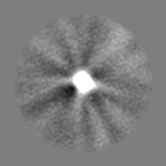
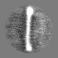
| Annotation | N.meningitidis NadAV3 homotrimeric map | ||||||||||||||||||||||||||||||||||||
| Projections & slices | Image control
Images are generated by Spider. | ||||||||||||||||||||||||||||||||||||
| Voxel size | X=Y=Z: 1.75 Å | ||||||||||||||||||||||||||||||||||||
| Density |
| ||||||||||||||||||||||||||||||||||||
| Symmetry | Space group: 1 | ||||||||||||||||||||||||||||||||||||
| Details | EMDB XML:
|
 Z (Sec.)
 Y (Row.)
 X (Col.)


-Supplemental data
-Half map: half1 map for FSC
| File | emd_19474_half_map_1.map | ||||||||||||
|---|---|---|---|---|---|---|---|---|---|---|---|---|---|
| Annotation | half1 map for FSC | ||||||||||||
| Projections & Slices |
| ||||||||||||
| Density Histograms |


-Half map: helf2 map for FSC
| File | emd_19474_half_map_2.map | ||||||||||||
|---|---|---|---|---|---|---|---|---|---|---|---|---|---|
| Annotation | helf2 map for FSC | ||||||||||||
| Projections & Slices |
| ||||||||||||
| Density Histograms |


- Sample components
-Entire : homotrimeric coiled-coil FL NadA bacterial adhesin expressed on t...
| Entire | Name: homotrimeric coiled-coil FL NadA bacterial adhesin expressed on the surface of bacteria N.meningitidis |
|---|---|
| Components |
|
-Supramolecule #1: homotrimeric coiled-coil FL NadA bacterial adhesin expressed on t...
| Supramolecule | Name: homotrimeric coiled-coil FL NadA bacterial adhesin expressed on the surface of bacteria N.meningitidis type: organelle_or_cellular_component / ID: 1 / Parent: 0 / Macromolecule list: all / Details: NadAV3 appendage generated |
|---|---|
| Source (natural) | Organism: Neisseria meningitidis (bacteria) / Strain: B |
-Macromolecule #1: NadAV3
| Macromolecule | Name: NadAV3 / type: other / ID: 1 / Classification: other |
|---|---|
| Source (natural) | Organism: Neisseria meningitidis serogroup B (bacteria) |
| Sequence | String: DDVKKAATVA IAAAYNNGQE INGFKAGETI YDIDEDGTIT KKDATAADVE ADDFKGLGL KKVVTNLTKT VNENKQNVDA KVKAAESEIE KLTTKLADTD A ALADTDAA LDATTNALNK LGENITTFAE ETKTNIVKID EKLEAASKHD DV KKAATVA IAAAYNNGQE ...String: DDVKKAATVA IAAAYNNGQE INGFKAGETI YDIDEDGTIT KKDATAADVE ADDFKGLGL KKVVTNLTKT VNENKQNVDA KVKAAESEIE KLTTKLADTD A ALADTDAA LDATTNALNK LGENITTFAE ETKTNIVKID EKLEAASKHD DV KKAATVA IAAAYNNGQE INGFKAGETI YDIDEDGTIT KKDATAADVE ADD FKGLGL KKVVTNLTKT VNENKQNVDA KVKAAESEIE KLTTKLADTD AALA DTDAA LDATTNALNK LGENITTFAE ETKTNIVKID EKLEAASDDV KKAAT VAIA AAYNNGQEIN GFKAGETIYD IDEDGTITKK DATAADVEAD DFKGLG LKK VVTNLTKTVN ENKQNVDAKV KAAESEIEKL TTKLADTDAA LADTDAA LD ATTNALNKLG ENITTFAEET KTNIVKIDEK LEAAS |
| Recombinant expression | Organism: |
-Experimental details
-Structure determination
| Method | cryo EM |
|---|---|
 Processing Processing | single particle reconstruction |
| Aggregation state | filament |
-Sample preparation
| Concentration | 1.00 mg/mL | |||||||||
|---|---|---|---|---|---|---|---|---|---|---|
| Buffer | pH: 7 Component:
Details: 20 mM Tris-HCL, 150 mM NaCl | |||||||||
| Grid | Model: EMS Lacey Carbon / Material: COPPER / Mesh: 400 / Support film - Material: CARBON / Support film - topology: HOLEY / Support film - Film thickness: 1 / Pretreatment - Type: GLOW DISCHARGE / Pretreatment - Time: 90 sec. / Pretreatment - Atmosphere: AIR Details: Quantifoil R2-2 grid, 400 mesh Cu, Electron Microscopy Sciences) was rendered hydrophilic with 15 mA current for 90 second by glow discharge in a EmiTech K100X | |||||||||
| Vitrification | Cryogen name: ETHANE / Chamber humidity: 100 % / Chamber temperature: 277.15 K / Instrument: FEI VITROBOT MARK IV Details: Quantifoil R2-2 grid, 400 mesh Cu, Electron Microscopy Sciences) charged with 2.5 microliter of the specimens were deposited onto the grid and vitrified using a Mark IV Vitrobot with a ...Details: Quantifoil R2-2 grid, 400 mesh Cu, Electron Microscopy Sciences) charged with 2.5 microliter of the specimens were deposited onto the grid and vitrified using a Mark IV Vitrobot with a blotting time of 4 second and humidity of 100 at 277.15K. | |||||||||
| Details | the sample was homogeneously distributed, no aggregation was observed. Original 1mg/ml NadAV3 sample was diluted up to 0.067 mg/ml for further EMM observations. |
- Electron microscopy
| Microscope | TFS KRIOS |
|---|---|
| Image recording | Film or detector model: FEI FALCON III (4k x 4k) / Average electron dose: 100.0 e/Å2 |
| Electron beam | Acceleration voltage: 300 kV / Electron source:  FIELD EMISSION GUN FIELD EMISSION GUN |
| Electron optics | Illumination mode: FLOOD BEAM / Imaging mode: BRIGHT FIELD / Nominal defocus max: 2.0 µm / Nominal defocus min: 0.4 µm |
| Experimental equipment |  Model: Titan Krios / Image courtesy: FEI Company |
+Image processing

-Atomic model buiding 1
| Initial model | PDB ID: Chain - Residue range: 24-170 / Chain - Source name: PDB / Chain - Initial model type: experimental model Details: the initial model consisted of the complete biological assembly for PDB entry 6EUN |
|---|---|
| Details | rigid-body fitting was done using ChimeraX. The position of rigid body fitted PDB 6EUN coordinates into the 3DEM map has been saved.Image of the rigid body fitting has been uploaded. |
| Refinement | Space: REAL / Protocol: RIGID BODY FIT / Overall B value: 0.4796 / Target criteria: cross-correlation coefficient |

