Grenoble Alliance for Integrated Structural Cell Biology (GRAL)
フランス
Grenoble Instruct-ERIC Center (ISBG)
フランス
Grenoble Instruct-ERIC Center (ISBG)
PID1697
フランス
Grenoble Instruct-ERIC Center (ISBG)
PID17035
フランス
European Regional Development Fund
UP CIISB
European Union
iNEXT-Discovery
MEYS CR 871037
European Union
Australian Research Council (ARC)
DP190100793
オーストラリア
引用
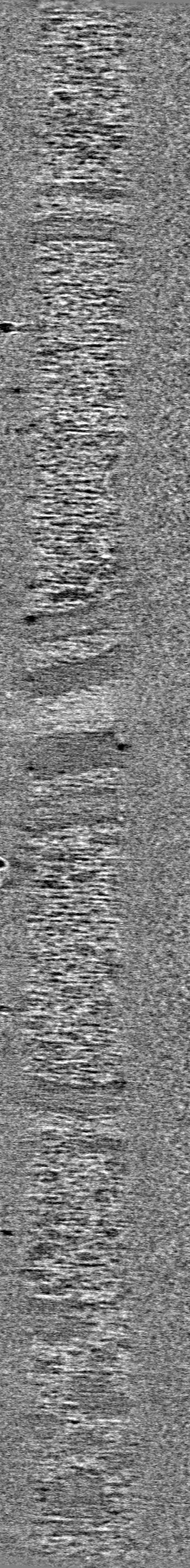
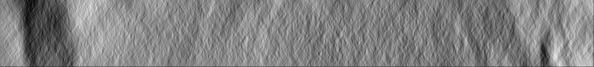
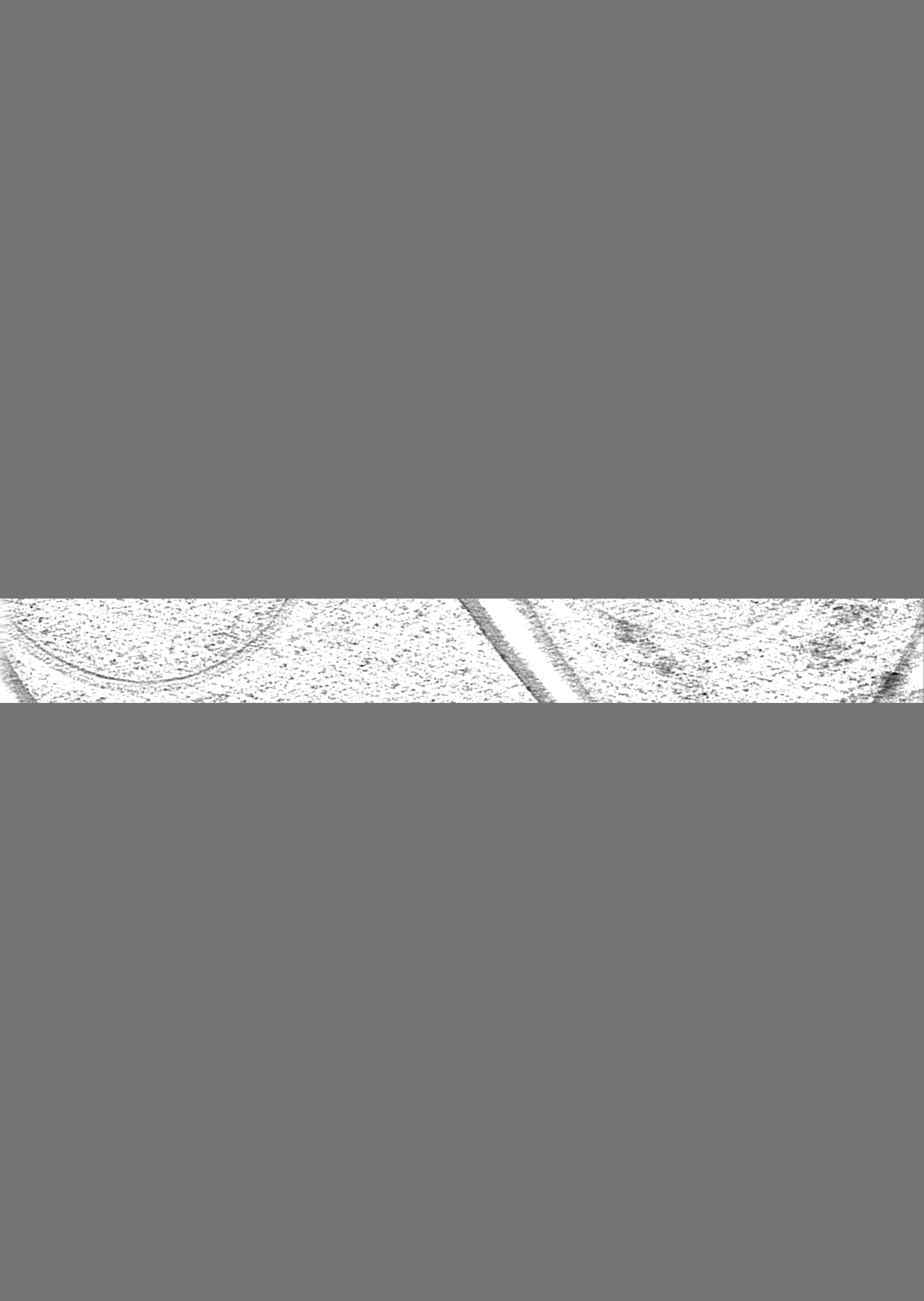
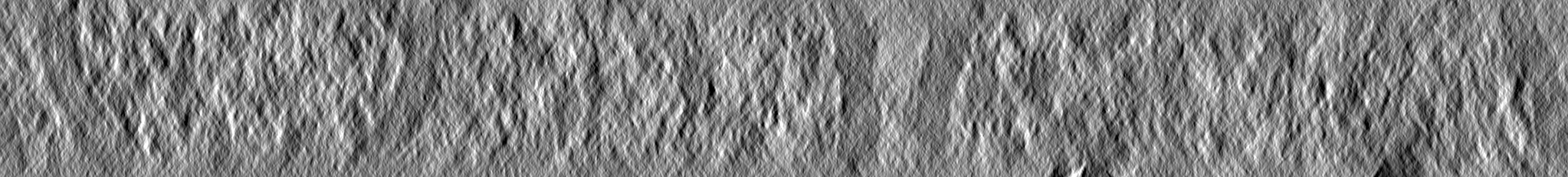
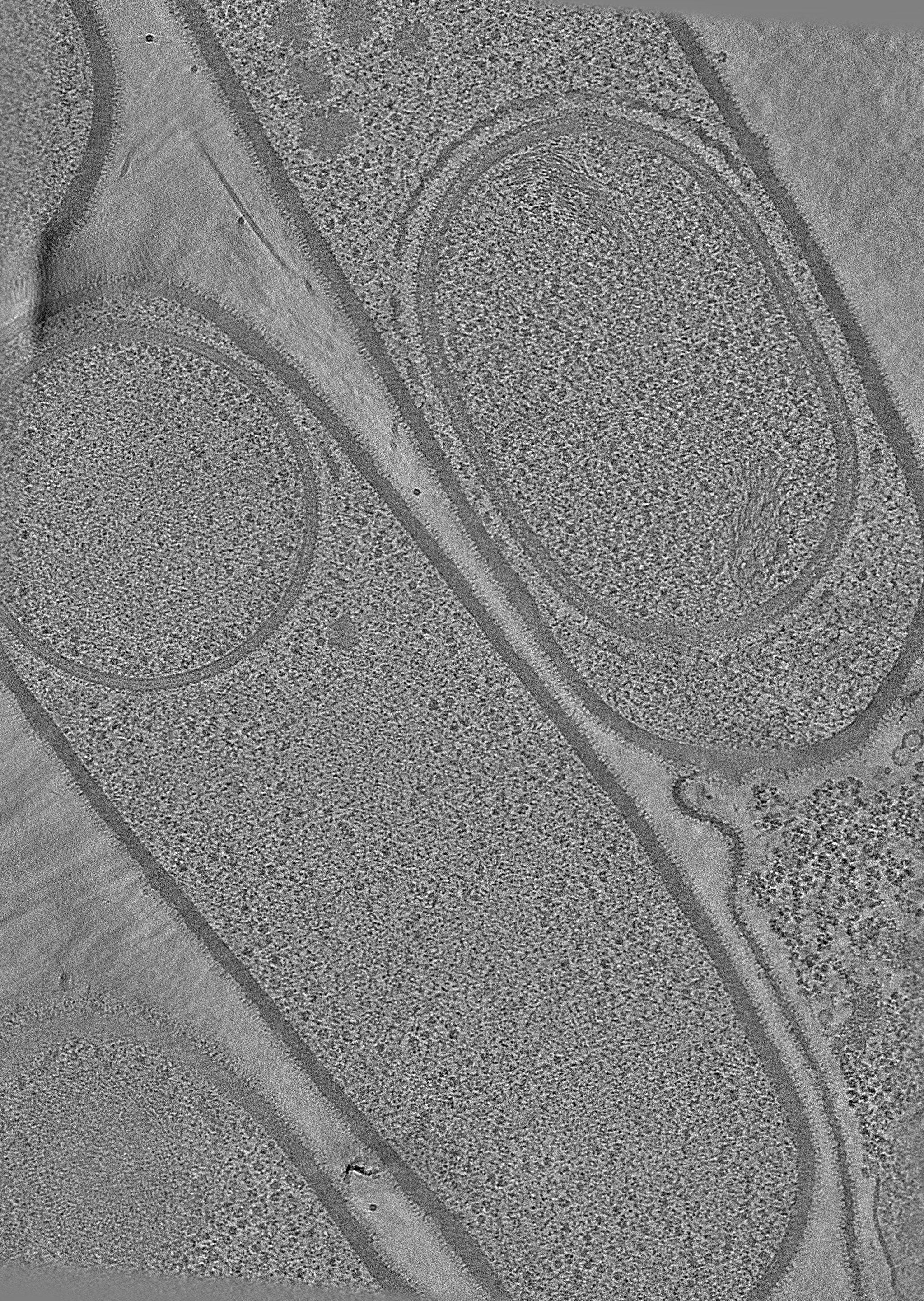
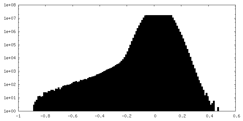
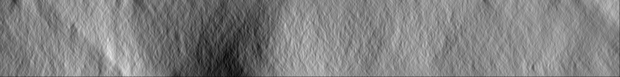
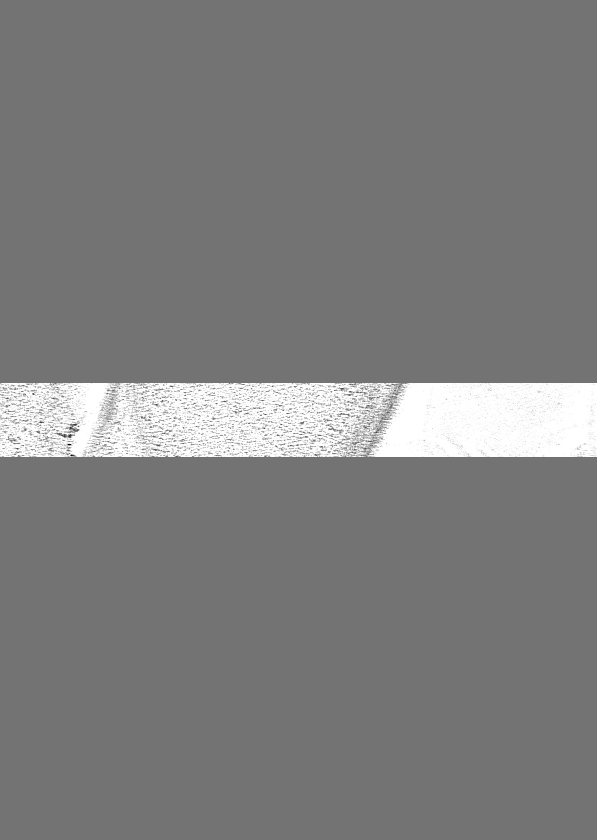
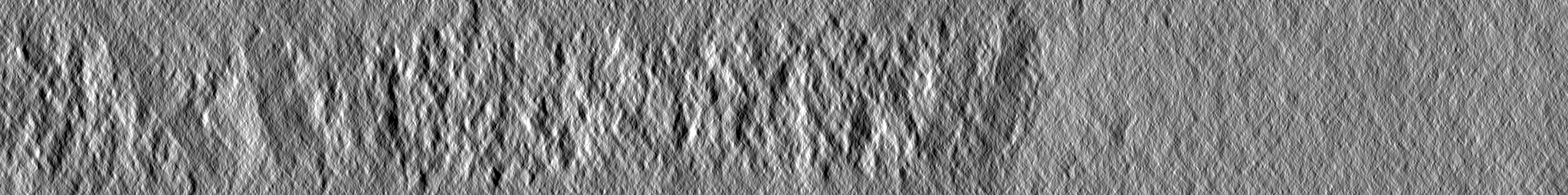
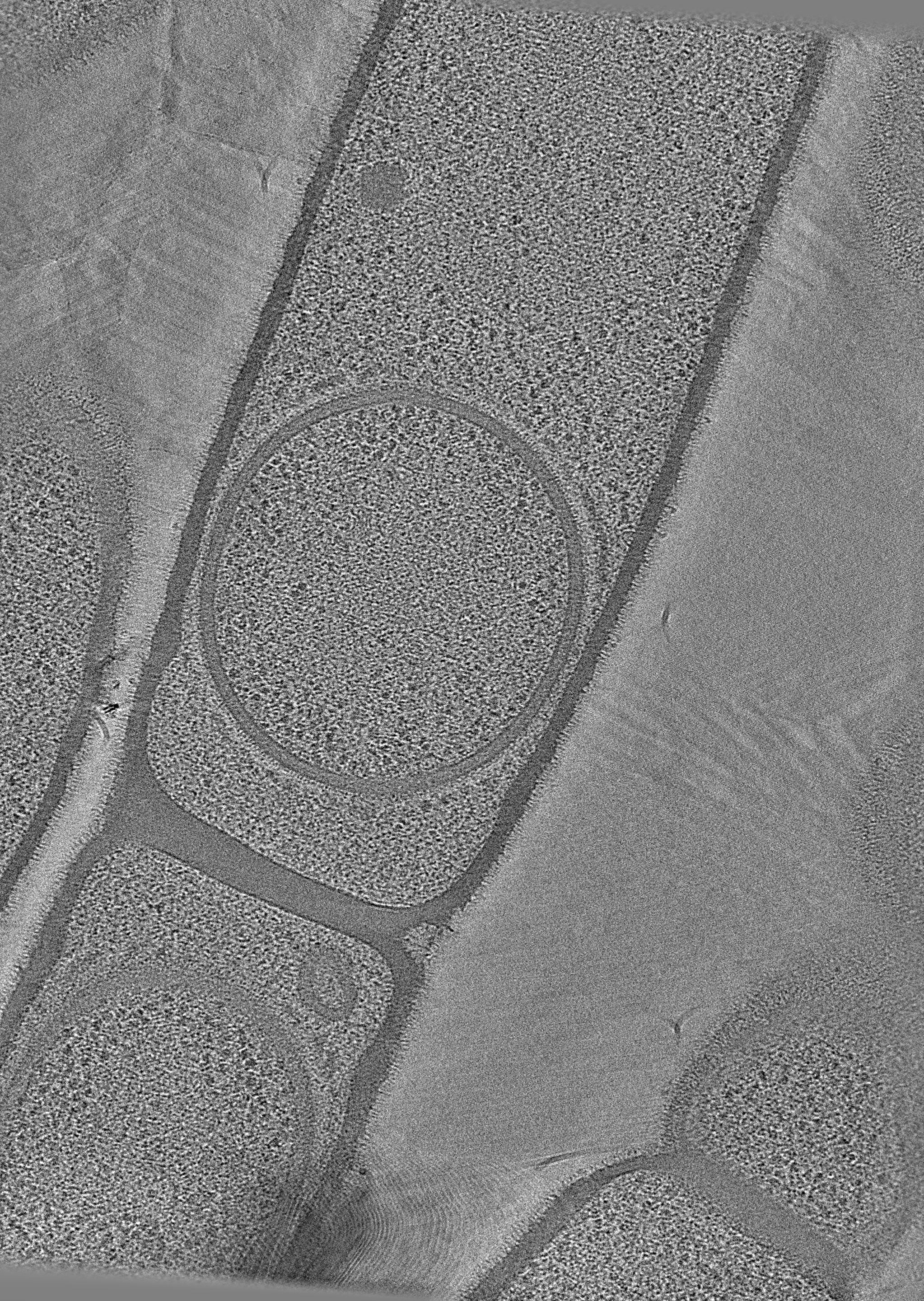
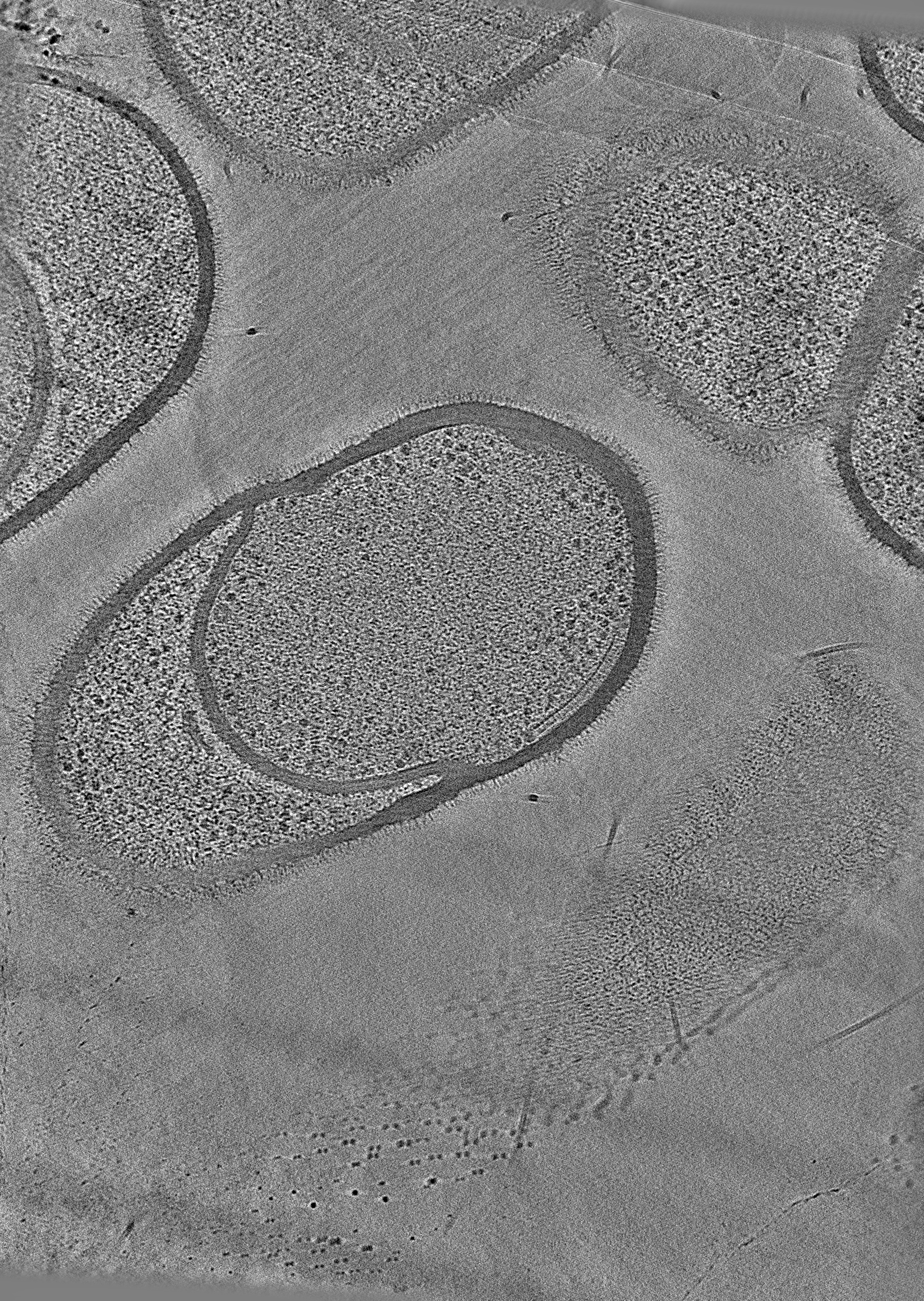
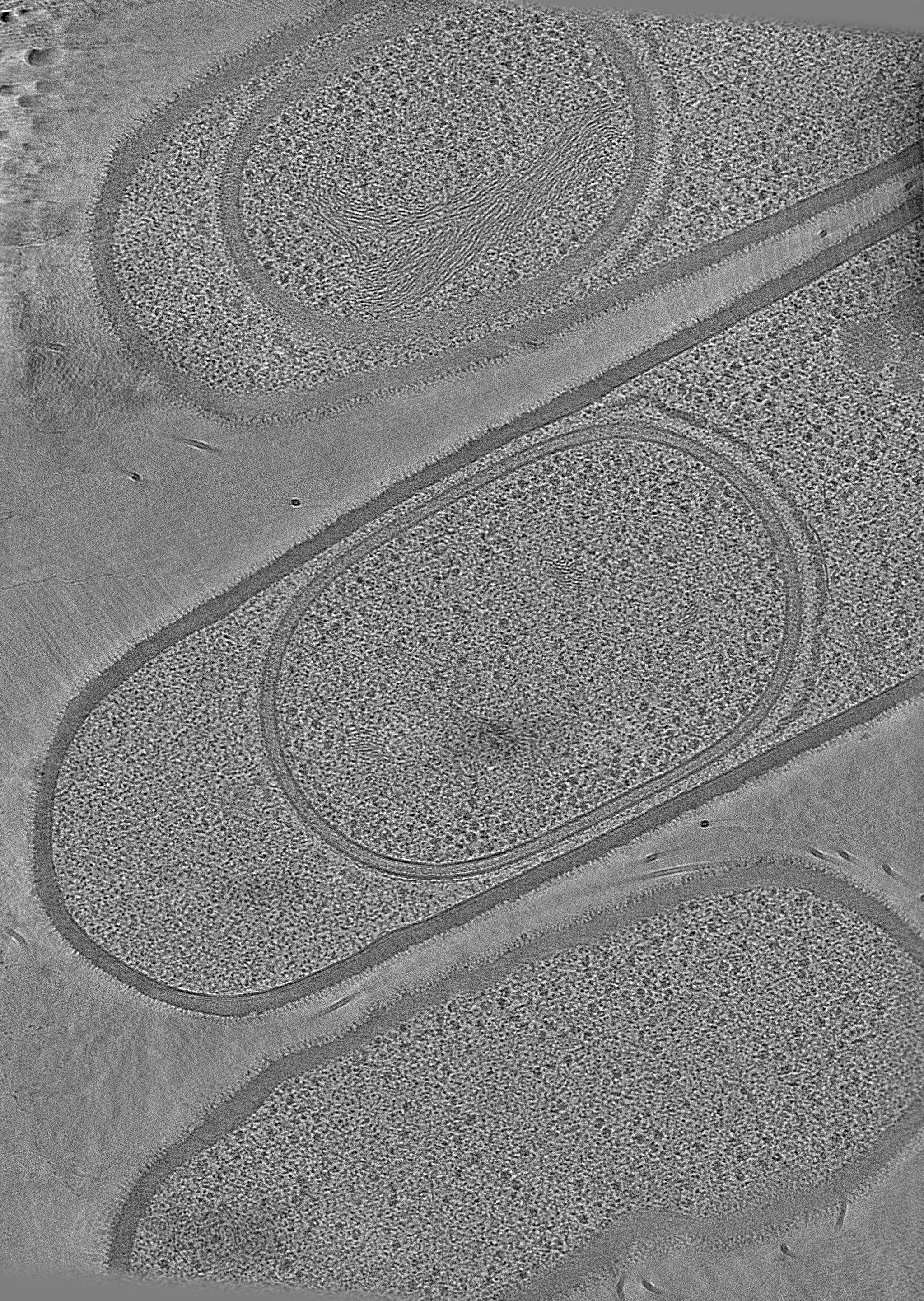
ジャーナル: Nat Commun / 年: 2024 タイトル: Ultrastructure of macromolecular assemblies contributing to bacterial spore resistance revealed by in situ cryo-electron tomography. 著者: Elda Bauda / Benoit Gallet / Jana Moravcova / Gregory Effantin / Helena Chan / Jiri Novacek / Pierre-Henri Jouneau / Christopher D A Rodrigues / Guy Schoehn / Christine Moriscot / Cecile Morlot / 要旨: Bacterial spores owe their incredible resistance capacities to molecular structures that protect the cell content from external aggressions. Among the determinants of resistance are the quaternary ...Bacterial spores owe their incredible resistance capacities to molecular structures that protect the cell content from external aggressions. Among the determinants of resistance are the quaternary structure of the chromosome and an extracellular shell made of proteinaceous layers (the coat), the assembly of which remains poorly understood. Here, in situ cryo-electron tomography on lamellae generated by cryo-focused ion beam micromachining provides insights into the ultrastructural organization of Bacillus subtilis sporangia. The reconstructed tomograms reveal that early during sporulation, the chromosome in the forespore adopts a toroidal structure harboring 5.5-nm thick fibers. At the same stage, coat proteins at the surface of the forespore form a stack of amorphous or structured layers with distinct electron density, dimensions and organization. By analyzing mutant strains using cryo-electron tomography and transmission electron microscopy on resin sections, we distinguish seven nascent coat regions with different molecular properties, and propose a model for the contribution of coat morphogenetic proteins.
全体 : Sporulating cells of Bacillus subtilis strain 168 deleted from th...
全体
名称: Sporulating cells of Bacillus subtilis strain 168 deleted from the cotE gene
要素
細胞: Sporulating cells of Bacillus subtilis strain 168 deleted from the cotE gene
-
超分子 #1: Sporulating cells of Bacillus subtilis strain 168 deleted from th...
超分子
名称: Sporulating cells of Bacillus subtilis strain 168 deleted from the cotE gene タイプ: cell / ID: 1 / 親要素: 0 詳細: View of a developing spore within a Bacillus subtilis mother cell
由来(天然)
生物種: Bacillus subtilis subsp. subtilis str. 168 (枯草菌)
-
実験情報
-
構造解析
手法
クライオ電子顕微鏡法
解析
電子線トモグラフィー法
試料の集合状態
cell
-
試料調製
緩衝液
pH: 7
凍結
凍結剤: ETHANE / チャンバー内湿度: 100 % / チャンバー内温度: 295 K / 装置: FEI VITROBOT MARK IV
Cryo protectant
5% glycerol
切片作成
集束イオンビーム - 装置: OTHER / 集束イオンビーム - イオン: OTHER / 集束イオンビーム - 電圧: 30 / 集束イオンビーム - 電流: 1 / 集束イオンビーム - 時間: 20 / 集束イオンビーム - 温度: 83 K / 集束イオンビーム - Initial thickness: 4000 / 集束イオンビーム - 最終 厚さ: 150 集束イオンビーム - 詳細: The value given for _em_focused_ion_beam.instrument is Zeiss Crossbeam 550. This is not in a list of allowed values {'DB235', 'OTHER'} so OTHER is written into the XML file.
-
電子顕微鏡法
顕微鏡
TFS KRIOS
撮影
フィルム・検出器のモデル: GATAN K3 BIOQUANTUM (6k x 4k) 平均電子線量: 85.0 e/Å2
 ムービー
ムービー コントローラー


 データを開く
 基本情報
基本情報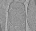
 マップデータ
 試料
試料 キーワード
キーワード
 データ登録者
 フランス, European Union,
  オーストラリア, 9件
  引用
引用

 構造の表示
構造の表示
 ダウンロードとリンク
 EMDBマップデータ形式
 emd_19411.png
 http://ftp.pdbj.org/pub/emdb/structures/EMD-19411
 Z
Z Y
Y X
X


 試料の構成要素
 解析
解析 電子顕微鏡法
 FIELD EMISSION GUN
