Grenoble Alliance for Integrated Structural Cell Biology (GRAL)
France
Grenoble Instruct-ERIC Center (ISBG)
France
Grenoble Instruct-ERIC Center (ISBG)
PID1697
France
Grenoble Instruct-ERIC Center (ISBG)
PID17035
France
European Regional Development Fund
UP CIISB
European Union
iNEXT-Discovery
MEYS CR 871037
European Union
Australian Research Council (ARC)
DP190100793
Australia
Citation
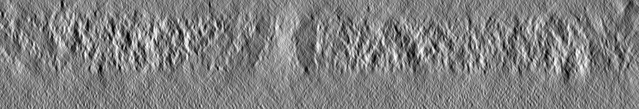
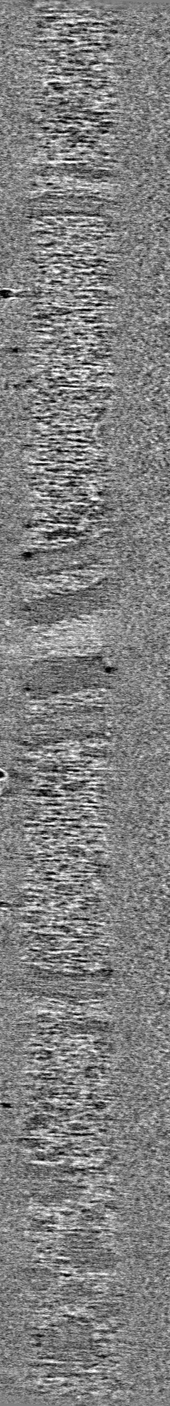
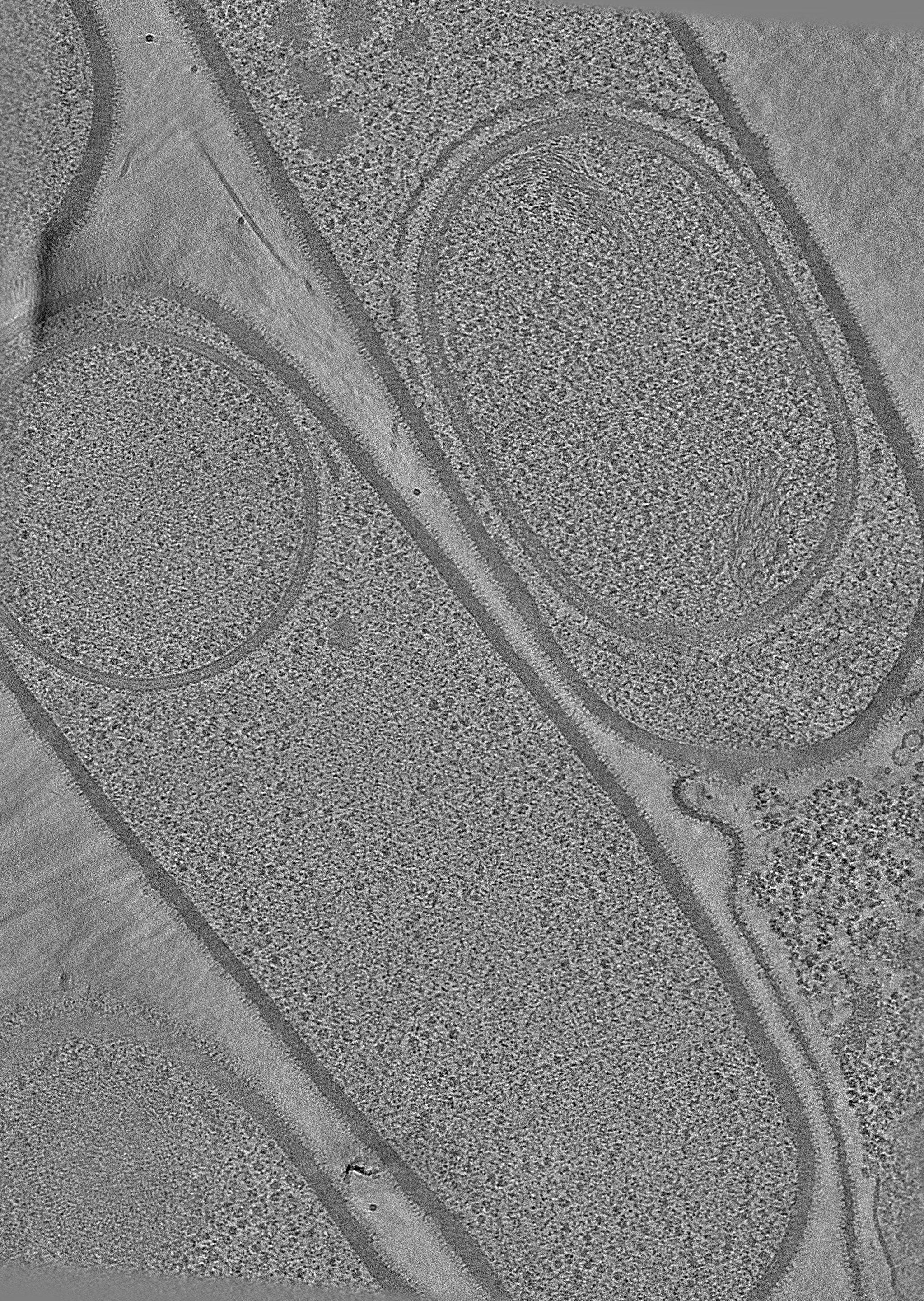
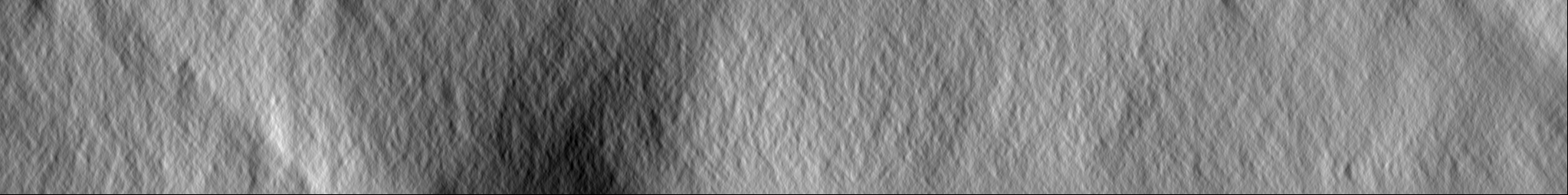
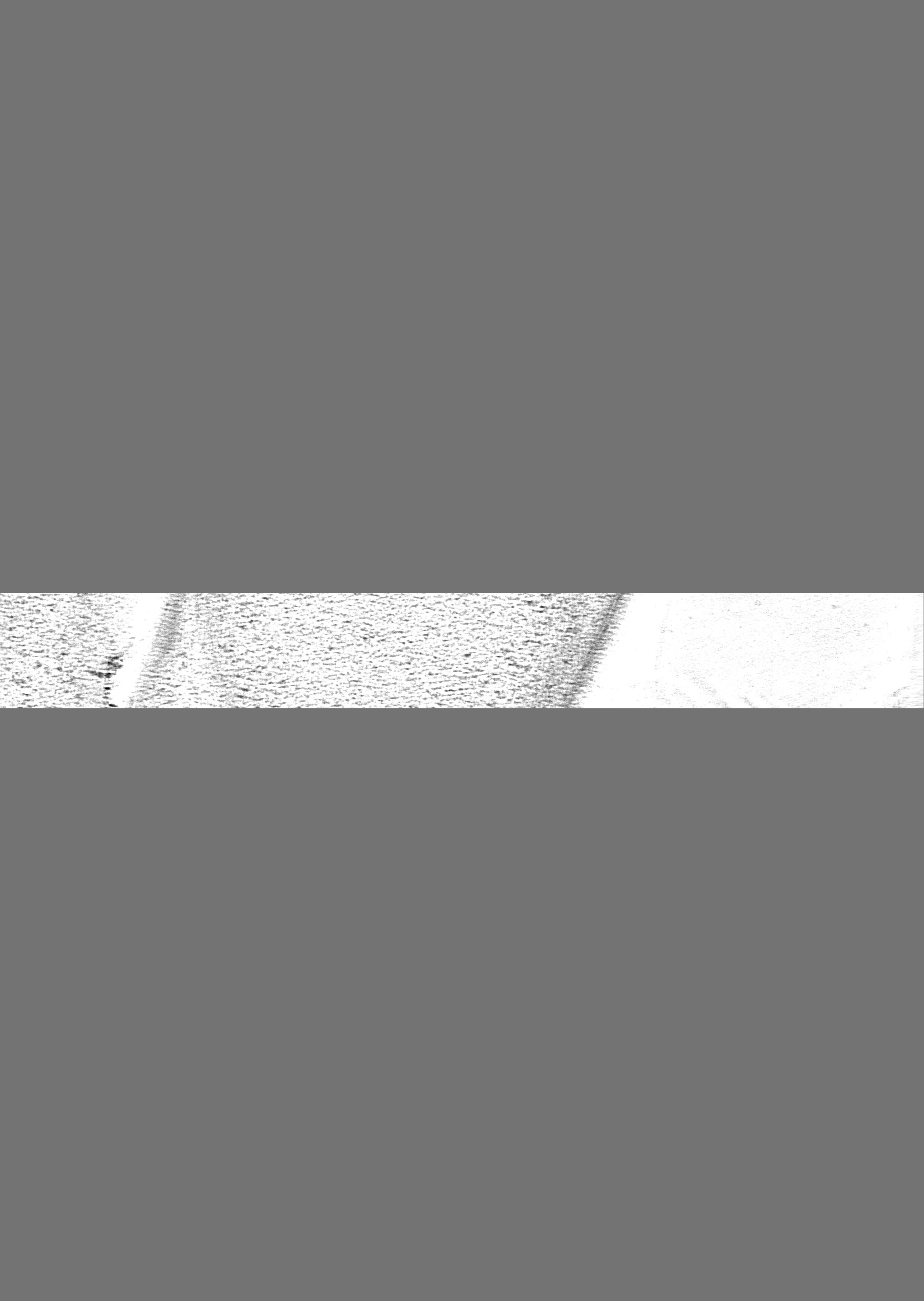
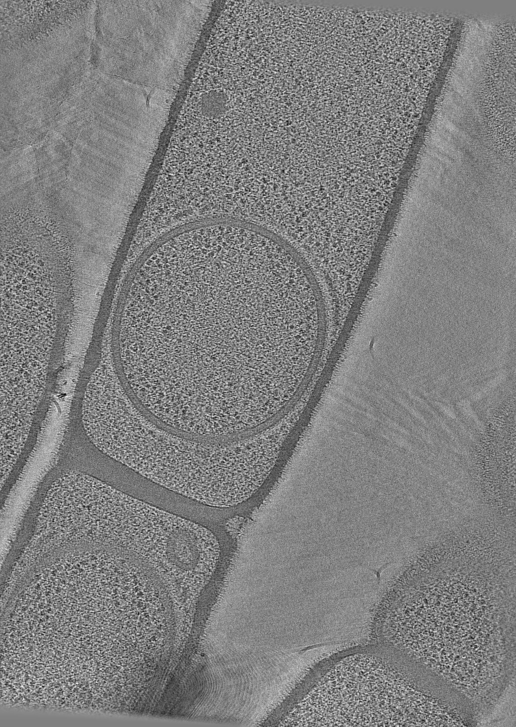
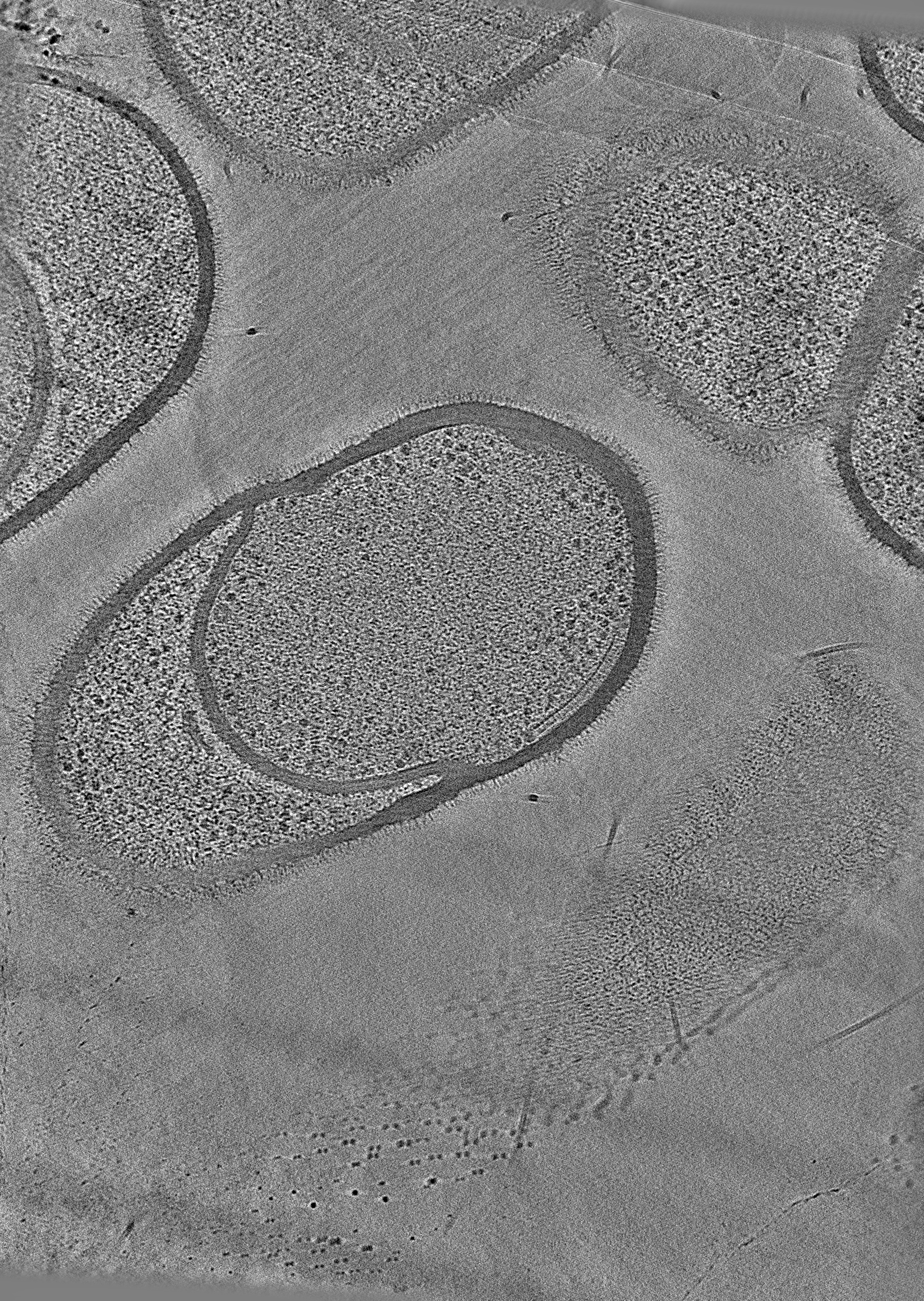
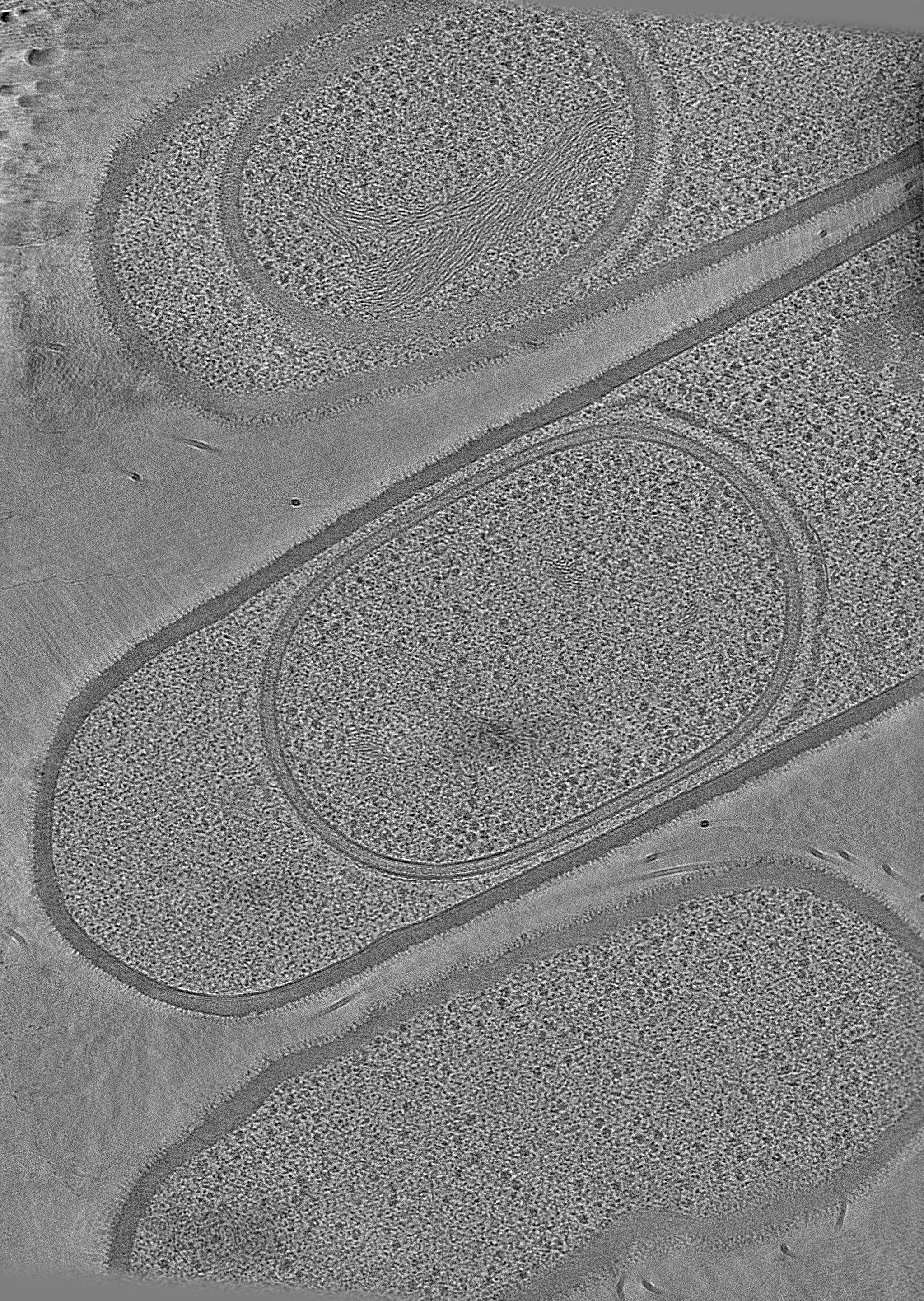
Journal: Nat Commun / Year: 2024 Title: Ultrastructure of macromolecular assemblies contributing to bacterial spore resistance revealed by in situ cryo-electron tomography. Authors: Elda Bauda / Benoit Gallet / Jana Moravcova / Gregory Effantin / Helena Chan / Jiri Novacek / Pierre-Henri Jouneau / Christopher D A Rodrigues / Guy Schoehn / Christine Moriscot / Cecile Morlot / Abstract: Bacterial spores owe their incredible resistance capacities to molecular structures that protect the cell content from external aggressions. Among the determinants of resistance are the quaternary ...Bacterial spores owe their incredible resistance capacities to molecular structures that protect the cell content from external aggressions. Among the determinants of resistance are the quaternary structure of the chromosome and an extracellular shell made of proteinaceous layers (the coat), the assembly of which remains poorly understood. Here, in situ cryo-electron tomography on lamellae generated by cryo-focused ion beam micromachining provides insights into the ultrastructural organization of Bacillus subtilis sporangia. The reconstructed tomograms reveal that early during sporulation, the chromosome in the forespore adopts a toroidal structure harboring 5.5-nm thick fibers. At the same stage, coat proteins at the surface of the forespore form a stack of amorphous or structured layers with distinct electron density, dimensions and organization. By analyzing mutant strains using cryo-electron tomography and transmission electron microscopy on resin sections, we distinguish seven nascent coat regions with different molecular properties, and propose a model for the contribution of coat morphogenetic proteins.
Entire : Sporulating cells of Bacillus subtilis strain 168 deleted from th...
Entire
Name: Sporulating cells of Bacillus subtilis strain 168 deleted from the cotE gene
Components
Cell: Sporulating cells of Bacillus subtilis strain 168 deleted from the cotE gene
-
Supramolecule #1: Sporulating cells of Bacillus subtilis strain 168 deleted from th...
Supramolecule
Name: Sporulating cells of Bacillus subtilis strain 168 deleted from the cotE gene type: cell / ID: 1 / Parent: 0 Details: View of a developing spore within a Bacillus subtilis mother cell
Source (natural)
Organism: Bacillus subtilis subsp. subtilis str. 168 (bacteria)
-
Experimental details
-
Structure determination
Method
cryo EM
Processing
electron tomography
Aggregation state
cell
-
Sample preparation
Buffer
pH: 7
Vitrification
Cryogen name: ETHANE / Chamber humidity: 100 % / Chamber temperature: 295 K / Instrument: FEI VITROBOT MARK IV
Cryo protectant
5% glycerol
Sectioning
Focused ion beam - Instrument: OTHER / Focused ion beam - Ion: OTHER / Focused ion beam - Voltage: 30 / Focused ion beam - Current: 1 / Focused ion beam - Duration: 20 / Focused ion beam - Temperature: 83 K / Focused ion beam - Initial thickness: 4000 / Focused ion beam - Final thickness: 150 Focused ion beam - Details: The value given for _em_focused_ion_beam.instrument is Zeiss Crossbeam 550. This is not in a list of allowed values {'DB235', 'OTHER'} so OTHER is written into the XML file.
-
Electron microscopy
Microscope
TFS KRIOS
Image recording
Film or detector model: GATAN K3 BIOQUANTUM (6k x 4k) / Average electron dose: 85.0 e/Å2
Electron beam
Acceleration voltage: 300 kV / Electron source: FIELD EMISSION GUN
In the structure databanks used in Yorodumi, some data are registered as the other names, "COVID-19 virus" and "2019-nCoV". Here are the details of the virus and the list of structure data.
Jan 31, 2019. EMDB accession codes are about to change! (news from PDBe EMDB page)
EMDB accession codes are about to change! (news from PDBe EMDB page)
The allocation of 4 digits for EMDB accession codes will soon come to an end. Whilst these codes will remain in use, new EMDB accession codes will include an additional digit and will expand incrementally as the available range of codes is exhausted. The current 4-digit format prefixed with “EMD-” (i.e. EMD-XXXX) will advance to a 5-digit format (i.e. EMD-XXXXX), and so on. It is currently estimated that the 4-digit codes will be depleted around Spring 2019, at which point the 5-digit format will come into force.
The EM Navigator/Yorodumi systems omit the EMD- prefix.
Related info.:Q: What is EMD? / ID/Accession-code notation in Yorodumi/EM Navigator
Yorodumi is a browser for structure data from EMDB, PDB, SASBDB, etc.
This page is also the successor to EM Navigator detail page, and also detail information page/front-end page for Omokage search.
The word "yorodu" (or yorozu) is an old Japanese word meaning "ten thousand". "mi" (miru) is to see.
Related info.:EMDB / PDB / SASBDB / Comparison of 3 databanks / Yorodumi Search / Aug 31, 2016. New EM Navigator & Yorodumi / Yorodumi Papers / Jmol/JSmol / Function and homology information / Changes in new EM Navigator and Yorodumi
 Movie
Movie Controller


 Open data
 Basic information
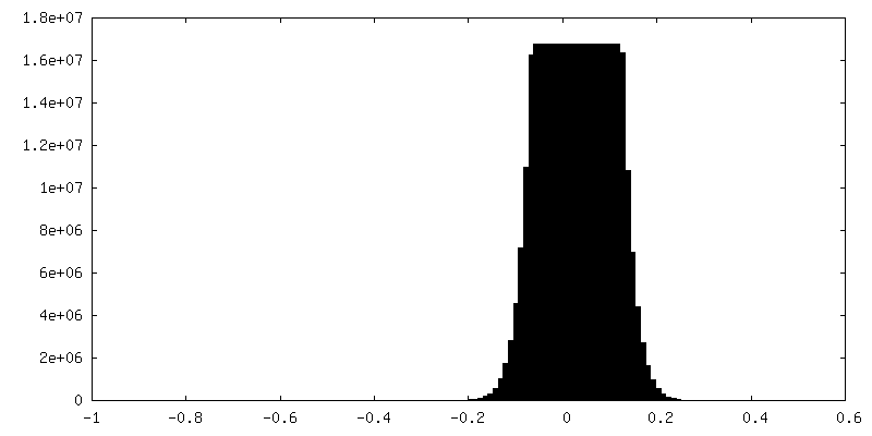
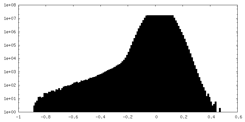
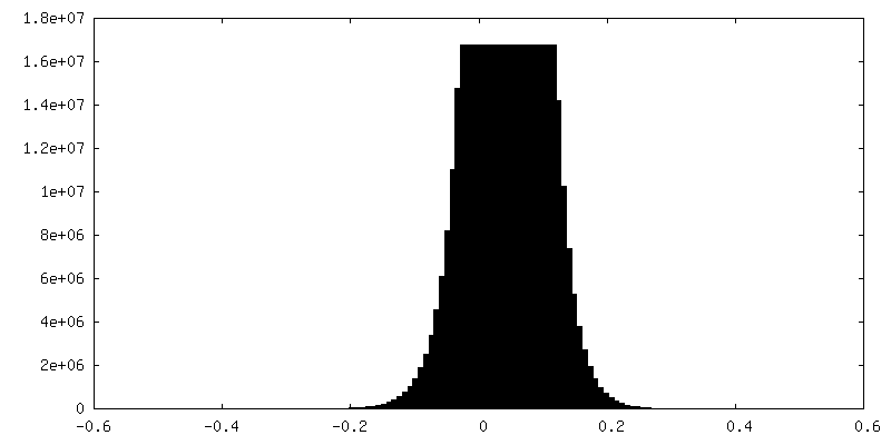
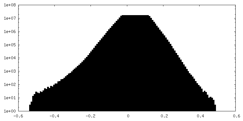
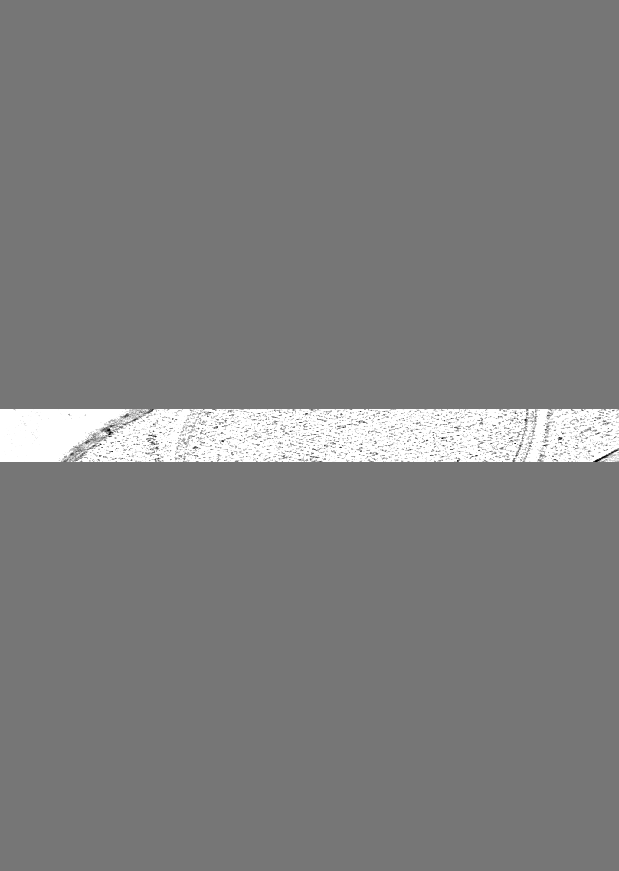
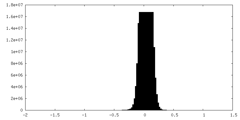
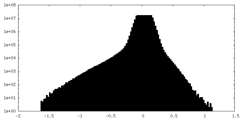
 Map data
 Sample
 Keywords
 Authors
 France, European Union,
  Australia, 9 items
  Citation

 Structure visualization
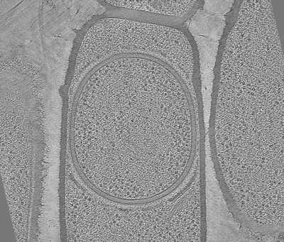
 Downloads & links
 EMDB map data format
 emd_19411.png
 http://ftp.pdbj.org/pub/emdb/structures/EMD-19411
 Z (Sec.)
 Y (Row.)
 X (Col.)


 Sample components
 Processing
 Electron microscopy
 FIELD EMISSION GUN
