- EMDB-19194: In situ cryo-electron tomogram of an autophagosome in the project... -
+
データを開く
IDまたはキーワード:
読み込み中...
-
基本情報
登録情報
データベース: EMDB / ID: EMD-19194
タイトル
In situ cryo-electron tomogram of an autophagosome in the projection of an iPSC-derived neuron #2
マップデータ
Tomogram of an autophagosome containing ER captured in the projection of an iPSC-derived neuron. Tomogram denoised with cryo-CARE with a model trained on this same tomogram.
試料
細胞器官・細胞要素: autophagosome containing membrane cargo captured in situ in the peripheral projections of iPSC-derived neurons grown a EM grid
キーワード
Autophagy / ERphagy / Degradation / Neuron / Cargo / Microtubules / ER / Autophagosome / CYTOSOLIC PROTEIN
ジャーナル: Nat Cell Biol / 年: 2024 タイトル: Combinatorial selective ER-phagy remodels the ER during neurogenesis. 著者: Melissa J Hoyer / Cristina Capitanio / Ian R Smith / Julia C Paoli / Anna Bieber / Yizhi Jiang / Joao A Paulo / Miguel A Gonzalez-Lozano / Wolfgang Baumeister / Florian Wilfling / Brenda A ...著者: Melissa J Hoyer / Cristina Capitanio / Ian R Smith / Julia C Paoli / Anna Bieber / Yizhi Jiang / Joao A Paulo / Miguel A Gonzalez-Lozano / Wolfgang Baumeister / Florian Wilfling / Brenda A Schulman / J Wade Harper / 要旨: The endoplasmic reticulum (ER) employs a diverse proteome landscape to orchestrate many cellular functions, ranging from protein and lipid synthesis to calcium ion flux and inter-organelle ...The endoplasmic reticulum (ER) employs a diverse proteome landscape to orchestrate many cellular functions, ranging from protein and lipid synthesis to calcium ion flux and inter-organelle communication. A case in point concerns the process of neurogenesis, where a refined tubular ER network is assembled via ER shaping proteins into the newly formed neuronal projections to create highly polarized dendrites and axons. Previous studies have suggested a role for autophagy in ER remodelling, as autophagy-deficient neurons in vivo display axonal ER accumulation within synaptic boutons, and the membrane-embedded ER-phagy receptor FAM134B has been genetically linked with human sensory and autonomic neuropathy. However, our understanding of the mechanisms underlying selective removal of the ER and the role of individual ER-phagy receptors is limited. Here we combine a genetically tractable induced neuron (iNeuron) system for monitoring ER remodelling during in vitro differentiation with proteomic and computational tools to create a quantitative landscape of ER proteome remodelling via selective autophagy. Through analysis of single and combinatorial ER-phagy receptor mutants, we delineate the extent to which each receptor contributes to both the magnitude and selectivity of ER protein clearance. We define specific subsets of ER membrane or lumenal proteins as preferred clients for distinct receptors. Using spatial sensors and flux reporters, we demonstrate receptor-specific autophagic capture of ER in axons, and directly visualize tubular ER membranes within autophagosomes in neuronal projections by cryo-electron tomography. This molecular inventory of ER proteome remodelling and versatile genetic toolkit provide a quantitative framework for understanding the contributions of individual ER-phagy receptors for reshaping ER during cell state transitions.
ダウンロード / ファイル: emd_19194.map.gz / 形式: CCP4 / 大きさ: 2.8 GB / タイプ: IMAGE STORED AS FLOATING POINT NUMBER (4 BYTES)
注釈
Tomogram of an autophagosome containing ER captured in the projection of an iPSC-derived neuron. Tomogram denoised with cryo-CARE with a model trained on this same tomogram.
ボクセルのサイズ
X=Y=Z: 11.72 Å
密度
最小 - 最大
-20.884499999999999 - 13.985004999999999
平均 (標準偏差)
0.15032952 (±0.8427676)
対称性
空間群: 1
詳細
EMDB XML:
マップ形状
Axis order
X
Y
Z
Origin
0
0
0
サイズ
1024
1024
712
Spacing
1024
1024
712
セル
A: 12001.28 Å / B: 12001.28 Å / C: 8344.641 Å α=β=γ: 90.0 °
-
添付データ
-
試料の構成要素
-
全体 : autophagosome containing membrane cargo captured in situ in the p...
全体
名称: autophagosome containing membrane cargo captured in situ in the peripheral projections of iPSC-derived neurons grown a EM grid
要素
細胞器官・細胞要素: autophagosome containing membrane cargo captured in situ in the peripheral projections of iPSC-derived neurons grown a EM grid
-
超分子 #1: autophagosome containing membrane cargo captured in situ in the p...
超分子
名称: autophagosome containing membrane cargo captured in situ in the peripheral projections of iPSC-derived neurons grown a EM grid タイプ: organelle_or_cellular_component / ID: 1 / 親要素: 0
由来(天然)
生物種: Homo sapiens (ヒト) / 株: iPSC KOLF2.0_AAVS-TREG3-NGN2 / 組織: induced-Neurons, DIV 18 / 細胞中の位置: neuronal projections
 ムービー
ムービー コントローラー


 データを開く
 基本情報
基本情報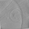
 マップデータ
 試料
試料 キーワード
キーワード Homo sapiens (ヒト)
 データ登録者
 米国, 1件
  引用
引用
 構造の表示
構造の表示
 ダウンロードとリンク
 EMDBマップデータ形式
 emd_19194.png
 http://ftp.pdbj.org/pub/emdb/structures/EMD-19194
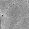
 試料の構成要素
 解析
解析 電子顕微鏡法
 FIELD EMISSION GUN
