Movie
Movie Controller


+ Open data

- Basic information
| Entry | Database: EMDB / ID: EMD-1909 | |||||||||
|---|---|---|---|---|---|---|---|---|---|---|
| Title | Skeletal thick filament showing myosin crowns and MyBP-C | |||||||||
 Map data Map data | Averaged thick filament tomogram showing myosin crowns and MyBP-C | |||||||||
 Sample Sample |
| |||||||||
 Keywords Keywords | C-protein / myosin binding protein C / electron tomography / thick filament structure | |||||||||
| Biological species | Anura (frogs & toads) | |||||||||
| Method | subtomogram averaging / cryo EM / Resolution: 70.0 Å | |||||||||
 Authors Authors | Luther PK / Winkler H / Taylor K / Zoghbi ME / Craig R / Padron R / Squire JM / Liu J | |||||||||
 Citation Citation | Journal: Proc Natl Acad Sci U S A / Year: 2011 Title: Direct visualization of myosin-binding protein C bridging myosin and actin filaments in intact muscle. Authors: Pradeep K Luther / Hanspeter Winkler / Kenneth Taylor / Maria E Zoghbi / Roger Craig / Raúl Padrón / John M Squire / Jun Liu /  Abstract: Myosin-binding protein C (MyBP-C) is a thick filament protein playing an essential role in muscle contraction, and MyBP-C mutations cause heart and skeletal muscle disease in millions worldwide. ...Myosin-binding protein C (MyBP-C) is a thick filament protein playing an essential role in muscle contraction, and MyBP-C mutations cause heart and skeletal muscle disease in millions worldwide. Despite its discovery 40 y ago, the mechanism of MyBP-C function remains unknown. In vitro studies suggest that MyBP-C could regulate contraction in a unique way--by bridging thick and thin filaments--but there has been no evidence for this in vivo. Here we use electron tomography of exceptionally well preserved muscle to demonstrate that MyBP-C does indeed bind to actin in intact muscle. This binding implies a physical mechanism for communicating the relative sliding between thick and thin filaments that does not involve myosin and which could modulate the contractile process. | |||||||||
| History |
|
- Structure visualization
| Movie |
 Movie viewer Movie viewer |
|---|---|
| Structure viewer | EM map: SurfViewMolmilJmol/JSmol |
| Supplemental images |

- Downloads & links
-EMDB archive
| Map data | emd_1909.map.gz | 12.6 MB | EMDB map data format | |
|---|---|---|---|---|
| Header (meta data) | emd-1909-v30.xmlemd-1909.xml | 7.5 KB 7.5 KB | Display Display | EMDB header |
| Images | 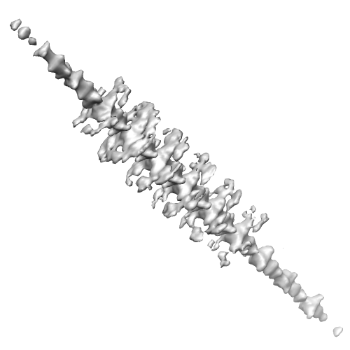 EMD-1909.png EMD-1909.png | 59 KB | ||
| Archive directory |  http://ftp.pdbj.org/pub/emdb/structures/EMD-1909ftp://ftp.pdbj.org/pub/emdb/structures/EMD-1909 http://ftp.pdbj.org/pub/emdb/structures/EMD-1909ftp://ftp.pdbj.org/pub/emdb/structures/EMD-1909 | HTTPS FTP |
-Links
| EMDB pages | EMDB (EBI/PDBe) / EMDataResource |
|---|
-Map
| File | Download / File: emd_1909.map.gz / Format: CCP4 / Size: 13.4 MB / Type: IMAGE STORED AS FLOATING POINT NUMBER (4 BYTES) | ||||||||||||||||||||||||||||||||||||||||||||||||||||||||||||||||||||
|---|---|---|---|---|---|---|---|---|---|---|---|---|---|---|---|---|---|---|---|---|---|---|---|---|---|---|---|---|---|---|---|---|---|---|---|---|---|---|---|---|---|---|---|---|---|---|---|---|---|---|---|---|---|---|---|---|---|---|---|---|---|---|---|---|---|---|---|---|---|
| Annotation | Averaged thick filament tomogram showing myosin crowns and MyBP-C | ||||||||||||||||||||||||||||||||||||||||||||||||||||||||||||||||||||
| Projections & slices | Image control
Images are generated by Spider. generated in cubic-lattice coordinate | ||||||||||||||||||||||||||||||||||||||||||||||||||||||||||||||||||||
| Voxel size | X=Y=Z: 11.5 Å | ||||||||||||||||||||||||||||||||||||||||||||||||||||||||||||||||||||
| Density |
| ||||||||||||||||||||||||||||||||||||||||||||||||||||||||||||||||||||
| Symmetry | Space group: 1 | ||||||||||||||||||||||||||||||||||||||||||||||||||||||||||||||||||||
| Details | EMDB XML:
CCP4 map header:
| ||||||||||||||||||||||||||||||||||||||||||||||||||||||||||||||||||||
 Z (Sec.)
 Y (Row.)
 X (Col.)


-Supplemental data
- Sample components
-Entire : Thin section of fast-frozen/freeze substituted relaxed frog sarto...
| Entire | Name: Thin section of fast-frozen/freeze substituted relaxed frog sartorius muscle. |
|---|---|
| Components |
|
-Supramolecule #1000: Thin section of fast-frozen/freeze substituted relaxed frog sarto...
| Supramolecule | Name: Thin section of fast-frozen/freeze substituted relaxed frog sartorius muscle. type: sample / ID: 1000 / Number unique components: 1 |
|---|---|
| Molecular weight | Experimental: 130 KDa / Theoretical: 130 KDa |
-Supramolecule #1: Thick filament
| Supramolecule | Name: Thick filament / type: organelle_or_cellular_component / ID: 1 / Name.synonym: Thick filament / Recombinant expression: No / Database: NCBI |
|---|---|
| Source (natural) | Organism: Anura (frogs & toads) |
-Experimental details
-Structure determination
| Method | cryo EM |
|---|---|
 Processing Processing | subtomogram averaging |
-Sample preparation
| Vitrification | Cryogen name: HELIUM / Instrument: OTHER |
|---|
- Electron microscopy
| Microscope | FEI/PHILIPS CM300FEG/T |
|---|---|
| Electron beam | Acceleration voltage: 300 kV / Electron source:  FIELD EMISSION GUN FIELD EMISSION GUN |
| Electron optics | Illumination mode: FLOOD BEAM / Imaging mode: BRIGHT FIELD |
| Sample stage | Specimen holder: Eucentric / Specimen holder model: OTHER |
-Image processing
| Final reconstruction | Algorithm: OTHER / Resolution.type: BY AUTHOR / Resolution: 70.0 Å / Resolution method: OTHER / Software - Name: Protomo |
|---|
-Atomic model buiding 1
| Initial model | PDB ID: |
|---|---|
| Refinement | Space: REAL |