 ムービー
ムービー コントローラー


+ データを開く

- 基本情報
基本情報
| 登録情報 | データベース: EMDB / ID: EMD-1906 | |||||||||
|---|---|---|---|---|---|---|---|---|---|---|
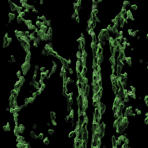
| タイトル | Tomographic reconstruction of unstained, frozen-hydrated thylakoid membranes from spinach chloroplasts. | |||||||||
 マップデータ マップデータ | Tomographic reconstruction of photosynthetic membranes from spinach chloroplasts resuspended in an isotonic buffer and imaged in an unstained frozen-hydrated state. | |||||||||
 試料 試料 |
| |||||||||
 キーワード キーワード | Photosynthesis / thylakoid membrane / CF1-CF0 proton ATPase / photosystem / cytochrome b6f / ribosome / chloroplast. | |||||||||
| 生物種 |  Spinacia oleracea (ホウレンソウ) Spinacia oleracea (ホウレンソウ) | |||||||||
| 手法 | 電子線トモグラフィー法 / クライオ電子顕微鏡法 / ネガティブ染色法 / 解像度: 45.0 Å | |||||||||
 データ登録者 データ登録者 | Ford RC / Holzenburg A | |||||||||
 引用 引用 | ジャーナル: Cystal Research and Technology / 年: 2013 タイトル: Organization of protein complexes and a mechanism for grana formation in photosynthetic membranes as revealed by cryo-electron microscopy 著者: Ford RC / Holzenburg A | |||||||||
| 履歴 |
|
- 構造の表示
構造の表示
| ムービー |
 ムービービューア ムービービューア |
|---|---|
| 構造ビューア | EMマップ: SurfViewMolmilJmol/JSmol |
| 添付画像 |
- ダウンロードとリンク
-EMDBアーカイブ
| マップデータ | emd_1906.map.gz | 149.7 MB | EMDBマップデータ形式 | |
|---|---|---|---|---|
| ヘッダ (付随情報) | emd-1906-v30.xmlemd-1906.xml | 10 KB 10 KB | 表示 表示 | EMDBヘッダ |
| 画像 | 1906.tif | 207.2 KB | ||
| アーカイブディレクトリ |  http://ftp.pdbj.org/pub/emdb/structures/EMD-1906ftp://ftp.pdbj.org/pub/emdb/structures/EMD-1906 http://ftp.pdbj.org/pub/emdb/structures/EMD-1906ftp://ftp.pdbj.org/pub/emdb/structures/EMD-1906 | HTTPS FTP |
-検証レポート
| 文書・要旨 | emd_1906_validation.pdf.gz | 180 KB | 表示 | EMDB検証レポート |
|---|---|---|---|---|
| 文書・詳細版 | emd_1906_full_validation.pdf.gz | 178.8 KB | 表示 | |
| XML形式データ | emd_1906_validation.xml.gz | 5.1 KB | 表示 | |
| アーカイブディレクトリ | https://ftp.pdbj.org/pub/emdb/validation_reports/EMD-1906ftp://ftp.pdbj.org/pub/emdb/validation_reports/EMD-1906 | HTTPS FTP |
-リンク
| EMDBのページ | EMDB (EBI/PDBe) / EMDataResource |
|---|
-マップ
| ファイル | ダウンロード / ファイル: emd_1906.map.gz / 形式: CCP4 / 大きさ: 294.5 MB / タイプ: IMAGE STORED AS SIGNED INTEGER (2 BYTES) | ||||||||||||||||||||||||||||||||||||||||||||||||||||||||||||||||||||
|---|---|---|---|---|---|---|---|---|---|---|---|---|---|---|---|---|---|---|---|---|---|---|---|---|---|---|---|---|---|---|---|---|---|---|---|---|---|---|---|---|---|---|---|---|---|---|---|---|---|---|---|---|---|---|---|---|---|---|---|---|---|---|---|---|---|---|---|---|---|
| 注釈 | Tomographic reconstruction of photosynthetic membranes from spinach chloroplasts resuspended in an isotonic buffer and imaged in an unstained frozen-hydrated state. | ||||||||||||||||||||||||||||||||||||||||||||||||||||||||||||||||||||
| 投影像・断面図 | 画像のコントロール
画像は Spider により作成 これらの図は立方格子座標系で作成されたものです | ||||||||||||||||||||||||||||||||||||||||||||||||||||||||||||||||||||
| ボクセルのサイズ | X=Y=Z: 17.6 Å | ||||||||||||||||||||||||||||||||||||||||||||||||||||||||||||||||||||
| 密度 |
| ||||||||||||||||||||||||||||||||||||||||||||||||||||||||||||||||||||
| 対称性 | 空間群: 1 | ||||||||||||||||||||||||||||||||||||||||||||||||||||||||||||||||||||
| 詳細 | EMDB XML:
CCP4マップ ヘッダ情報:
| ||||||||||||||||||||||||||||||||||||||||||||||||||||||||||||||||||||
 Z (Sec.)
 Y (Row.)
 X (Col.)


-添付データ
- 試料の構成要素
-全体 : Thylakoid membrane from spinach.
| 全体 | 名称: Thylakoid membrane from spinach. |
|---|---|
| 要素 |
|
-超分子 #1000: Thylakoid membrane from spinach.
| 超分子 | 名称: Thylakoid membrane from spinach. / タイプ: sample / ID: 1000 / Number unique components: 1 |
|---|
-超分子 #1: Thylakoid membrane
| 超分子 | 名称: Thylakoid membrane / タイプ: organelle_or_cellular_component / ID: 1 / Name.synonym: Photosynthetic membrane 詳細: Buffer - 20mM MES, 5mM MgCl2, 15mM NaCl, pH6.5, 150mM sorbitol. 3microlitres of membranes at 2mg per ml chlorophyll were directly applied to electron microscope grids (Quantifoil carbon grids ...詳細: Buffer - 20mM MES, 5mM MgCl2, 15mM NaCl, pH6.5, 150mM sorbitol. 3microlitres of membranes at 2mg per ml chlorophyll were directly applied to electron microscope grids (Quantifoil carbon grids with 1.2 micron holes). After blotting excess liquid, (2x 1s blots at 95 percent relative humidity in a Vitrobot device), the grids were rapidly frozen by plunging into liquid ethane and transferred at less than 110K to the Gatan cryo-stage of the electron microscope. 組換発現: No / データベース: NCBI |
|---|---|
| 由来(天然) | 生物種: Spinacia oleracea (ホウレンソウ) / 別称: Spinach / 組織: Leaf / Organelle: Chloroplast |
-実験情報
-構造解析
| 手法 | ネガティブ染色法, クライオ電子顕微鏡法 |
|---|---|
 解析 解析 | 電子線トモグラフィー法 |
-試料調製
| 濃度 | 2 mg/mL |
|---|---|
| 緩衝液 | pH: 6.5 詳細: 20mM MES, 5mM MgCl2, 15mM NaCl, pH6.5, 150mM sorbitol. |
| 染色 | タイプ: NEGATIVE / 詳細: No stain. |
| グリッド | 詳細: Quantifoil 400mesh carbon grids with 1.2 micron holes. |
| 凍結 | 凍結剤: NITROGEN / チャンバー内湿度: 95 % / チャンバー内温度: 97 K / 装置: OTHER 詳細: Vitrification instrument: Vitrobot FEI. Rapid plunging into liquid ethane. 手法: 2 x 1 sec blots before plunging. |
- 電子顕微鏡法
| 顕微鏡 | FEI TECNAI F20 |
|---|---|
| 温度 | 最低: 96 K / 最高: 98 K / 平均: 97 K |
| アライメント法 | Legacy - 非点収差: Astigmatism corrected by live FFT. |
| 撮影 | デジタル化 - サンプリング間隔: 15 µm / 実像数: 57 / 平均電子線量: 45 e/Å2 |
| 電子線 | 加速電圧: 200 kV / 電子線源:  FIELD EMISSION GUN FIELD EMISSION GUN |
| 電子光学系 | 倍率(補正後): 33998 / 照射モード: FLOOD BEAM / 撮影モード: BRIGHT FIELD / Cs: 2.0 mm / 最大 デフォーカス(公称値): 8.0 µm / 最小 デフォーカス(公称値): 8.0 µm / 倍率(公称値): 34000 |
| 試料ステージ | 試料ホルダー: Single tilt / 試料ホルダーモデル: GATAN LIQUID NITROGEN / Tilt series - Axis1 - Min angle: -65 ° / Tilt series - Axis1 - Max angle: 49 ° / Tilt series - Axis1 - Angle increment: 1 ° |
| 実験機器 |  モデル: Tecnai F20 / 画像提供: FEI Company |
-画像解析
| 詳細 | 1 degree increments above 45 deg, 2.5 degree increments below this. |
|---|---|
| 最終 再構成 | 解像度のタイプ: BY AUTHOR / 解像度: 45.0 Å / 解像度の算出法: OTHER / ソフトウェア - 名称: eTOMO 詳細: Standard eTOMO procedures for single axis tilt tomograms. Fiducials were selected from the tomogram and tracked in each image by hand initially. 使用した粒子像数: 57 |
| CTF補正 | 詳細: none |
