 Movie
Movie Controller

[English] 日本語
 Yorodumi
- EMDB-1906: Tomographic reconstruction of unstained, frozen-hydrated thylakoi... -
+ Open data

- Basic information
| Entry | Database: EMDB / ID: EMD-1906 | |||||||||
|---|---|---|---|---|---|---|---|---|---|---|
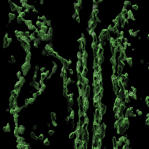
| Title | Tomographic reconstruction of unstained, frozen-hydrated thylakoid membranes from spinach chloroplasts. | |||||||||
 Map data Map data | Tomographic reconstruction of photosynthetic membranes from spinach chloroplasts resuspended in an isotonic buffer and imaged in an unstained frozen-hydrated state. | |||||||||
 Sample Sample |
| |||||||||
 Keywords Keywords | Photosynthesis / thylakoid membrane / CF1-CF0 proton ATPase / photosystem / cytochrome b6f / ribosome / chloroplast. | |||||||||
| Biological species |  Spinacia oleracea (spinach) Spinacia oleracea (spinach) | |||||||||
| Method | electron tomography / cryo EM / negative staining / Resolution: 45.0 Å | |||||||||
 Authors Authors | Ford RC / Holzenburg A | |||||||||
 Citation Citation | Journal: Cystal Research and Technology / Year: 2013 Title: Organization of protein complexes and a mechanism for grana formation in photosynthetic membranes as revealed by cryo-electron microscopy Authors: Ford RC / Holzenburg A | |||||||||
| History |
|
- Structure visualization
| Movie |
 Movie viewer Movie viewer |
|---|---|
| Structure viewer | EM map: SurfViewMolmilJmol/JSmol |
| Supplemental images |
- Downloads & links
-EMDB archive
| Map data | emd_1906.map.gz | 149.7 MB | EMDB map data format | |
|---|---|---|---|---|
| Header (meta data) | emd-1906-v30.xmlemd-1906.xml | 10 KB 10 KB | Display Display | EMDB header |
| Images | 1906.tif | 207.2 KB | ||
| Archive directory |  http://ftp.pdbj.org/pub/emdb/structures/EMD-1906ftp://ftp.pdbj.org/pub/emdb/structures/EMD-1906 http://ftp.pdbj.org/pub/emdb/structures/EMD-1906ftp://ftp.pdbj.org/pub/emdb/structures/EMD-1906 | HTTPS FTP |
-Validation report
| Summary document | emd_1906_validation.pdf.gz | 180 KB | Display | EMDB validaton report |
|---|---|---|---|---|
| Full document | emd_1906_full_validation.pdf.gz | 178.8 KB | Display | |
| Data in XML | emd_1906_validation.xml.gz | 5.1 KB | Display | |
| Arichive directory | https://ftp.pdbj.org/pub/emdb/validation_reports/EMD-1906ftp://ftp.pdbj.org/pub/emdb/validation_reports/EMD-1906 | HTTPS FTP |
-Links
| EMDB pages | EMDB (EBI/PDBe) / EMDataResource |
|---|
-Map
| File | Download / File: emd_1906.map.gz / Format: CCP4 / Size: 294.5 MB / Type: IMAGE STORED AS SIGNED INTEGER (2 BYTES) | ||||||||||||||||||||||||||||||||||||||||||||||||||||||||||||||||||||
|---|---|---|---|---|---|---|---|---|---|---|---|---|---|---|---|---|---|---|---|---|---|---|---|---|---|---|---|---|---|---|---|---|---|---|---|---|---|---|---|---|---|---|---|---|---|---|---|---|---|---|---|---|---|---|---|---|---|---|---|---|---|---|---|---|---|---|---|---|---|
| Annotation | Tomographic reconstruction of photosynthetic membranes from spinach chloroplasts resuspended in an isotonic buffer and imaged in an unstained frozen-hydrated state. | ||||||||||||||||||||||||||||||||||||||||||||||||||||||||||||||||||||
| Projections & slices | Image control
Images are generated by Spider. generated in cubic-lattice coordinate | ||||||||||||||||||||||||||||||||||||||||||||||||||||||||||||||||||||
| Voxel size | X=Y=Z: 17.6 Å | ||||||||||||||||||||||||||||||||||||||||||||||||||||||||||||||||||||
| Density |
| ||||||||||||||||||||||||||||||||||||||||||||||||||||||||||||||||||||
| Symmetry | Space group: 1 | ||||||||||||||||||||||||||||||||||||||||||||||||||||||||||||||||||||
| Details | EMDB XML:
CCP4 map header:
| ||||||||||||||||||||||||||||||||||||||||||||||||||||||||||||||||||||
 Z (Sec.)
 Y (Row.)
 X (Col.)


-Supplemental data
- Sample components
-Entire : Thylakoid membrane from spinach.
| Entire | Name: Thylakoid membrane from spinach. |
|---|---|
| Components |
|
-Supramolecule #1000: Thylakoid membrane from spinach.
| Supramolecule | Name: Thylakoid membrane from spinach. / type: sample / ID: 1000 / Number unique components: 1 |
|---|
-Supramolecule #1: Thylakoid membrane
| Supramolecule | Name: Thylakoid membrane / type: organelle_or_cellular_component / ID: 1 / Name.synonym: Photosynthetic membrane Details: Buffer - 20mM MES, 5mM MgCl2, 15mM NaCl, pH6.5, 150mM sorbitol. 3microlitres of membranes at 2mg per ml chlorophyll were directly applied to electron microscope grids (Quantifoil carbon ...Details: Buffer - 20mM MES, 5mM MgCl2, 15mM NaCl, pH6.5, 150mM sorbitol. 3microlitres of membranes at 2mg per ml chlorophyll were directly applied to electron microscope grids (Quantifoil carbon grids with 1.2 micron holes). After blotting excess liquid, (2x 1s blots at 95 percent relative humidity in a Vitrobot device), the grids were rapidly frozen by plunging into liquid ethane and transferred at less than 110K to the Gatan cryo-stage of the electron microscope. Recombinant expression: No / Database: NCBI |
|---|---|
| Source (natural) | Organism: Spinacia oleracea (spinach) / synonym: Spinach / Tissue: Leaf / Organelle: Chloroplast |
-Experimental details
-Structure determination
| Method | negative staining, cryo EM |
|---|---|
 Processing Processing | electron tomography |
-Sample preparation
| Concentration | 2 mg/mL |
|---|---|
| Buffer | pH: 6.5 Details: 20mM MES, 5mM MgCl2, 15mM NaCl, pH6.5, 150mM sorbitol. |
| Staining | Type: NEGATIVE / Details: No stain. |
| Grid | Details: Quantifoil 400mesh carbon grids with 1.2 micron holes. |
| Vitrification | Cryogen name: NITROGEN / Chamber humidity: 95 % / Chamber temperature: 97 K / Instrument: OTHER Details: Vitrification instrument: Vitrobot FEI. Rapid plunging into liquid ethane. Method: 2 x 1 sec blots before plunging. |
- Electron microscopy
| Microscope | FEI TECNAI F20 |
|---|---|
| Temperature | Min: 96 K / Max: 98 K / Average: 97 K |
| Alignment procedure | Legacy - Astigmatism: Astigmatism corrected by live FFT. |
| Image recording | Digitization - Sampling interval: 15 µm / Number real images: 57 / Average electron dose: 45 e/Å2 |
| Electron beam | Acceleration voltage: 200 kV / Electron source:  FIELD EMISSION GUN FIELD EMISSION GUN |
| Electron optics | Calibrated magnification: 33998 / Illumination mode: FLOOD BEAM / Imaging mode: BRIGHT FIELD / Cs: 2.0 mm / Nominal defocus max: 8.0 µm / Nominal defocus min: 8.0 µm / Nominal magnification: 34000 |
| Sample stage | Specimen holder: Single tilt / Specimen holder model: GATAN LIQUID NITROGEN / Tilt series - Axis1 - Min angle: -65 ° / Tilt series - Axis1 - Max angle: 49 ° / Tilt series - Axis1 - Angle increment: 1 ° |
| Experimental equipment |  Model: Tecnai F20 / Image courtesy: FEI Company |
-Image processing
| Details | 1 degree increments above 45 deg, 2.5 degree increments below this. |
|---|---|
| Final reconstruction | Resolution.type: BY AUTHOR / Resolution: 45.0 Å / Resolution method: OTHER / Software - Name: eTOMO Details: Standard eTOMO procedures for single axis tilt tomograms. Fiducials were selected from the tomogram and tracked in each image by hand initially. Number images used: 57 |
| CTF correction | Details: none |
