 ムービー
ムービー コントローラー


+ データを開く

- 基本情報
基本情報
| 登録情報 | データベース: EMDB / ID: EMD-1904 | |||||||||
|---|---|---|---|---|---|---|---|---|---|---|
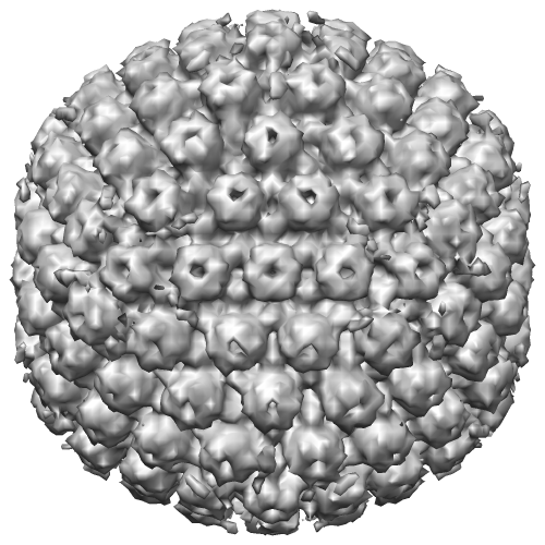
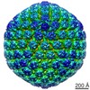
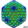
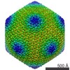
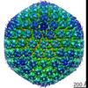
| タイトル | Labeling and Localization of the Herpes Simplex Virus Capsid Protein UL25 and Its Interaction with the Two Triplexes Closest to the Penton. | |||||||||
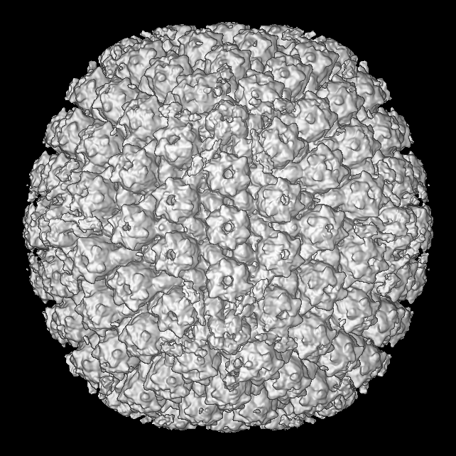
 マップデータ マップデータ | Surface rendered view of HSV-1 C-capsid where protein UL25 has been genetically tagged with GFP at amino acid 50. | |||||||||
 試料 試料 |
| |||||||||
 キーワード キーワード | HSV-1 / capsid / UL25 / cryo-EM | |||||||||
| 生物種 |  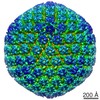 Human herpesvirus 1 strain KOS (ヘルペスウイルス) Human herpesvirus 1 strain KOS (ヘルペスウイルス) | |||||||||
| 手法 | 単粒子再構成法 / クライオ電子顕微鏡法 / 解像度: 13.7 Å | |||||||||
 データ登録者 データ登録者 | Conway JF / Cockrell SK / Copeland AM / Newcomb WW / Brown JC / Homa FL | |||||||||
 引用 引用 | ジャーナル: J Mol Biol / 年: 2010 タイトル: Labeling and localization of the herpes simplex virus capsid protein UL25 and its interaction with the two triplexes closest to the penton. 著者: James F Conway / Shelley K Cockrell / Anna Maria Copeland / William W Newcomb / Jay C Brown / Fred L Homa /  要旨: The herpes simplex virus type 1 UL25 protein is one of seven viral proteins that are required for DNA cleavage and packaging. Together with UL17, UL25 forms part of an elongated molecule referred to ...The herpes simplex virus type 1 UL25 protein is one of seven viral proteins that are required for DNA cleavage and packaging. Together with UL17, UL25 forms part of an elongated molecule referred to as the C-capsid-specific component (CCSC). Five copies of the CCSC are located at each of the capsid vertices on DNA-containing capsids. To study the conformation of UL25 as it is folded on the capsid surface, we identified the sequence recognized by a UL25-specific monoclonal antibody and localized the epitope on the capsid surface by immunogold electron microscopy. The epitope mapped to amino acids 99-111 adjacent to the region of the protein (amino acids 1-50) that is required for capsid binding. In addition, cryo-EM reconstructions of C-capsids in which the green fluorescent protein (GFP) was fused within the N-terminus of UL25 localized the point of contact between UL25 and GFP. The result confirmed the modeled location of the UL25 protein in the CCSC density as the region that is distal to the penton with the N-terminus of UL25 making contact with the triplex one removed from the penton. Immunofluorescence experiments at early times during infection demonstrated that UL25-GFP was present on capsids located within the cytoplasm and adjacent to the nucleus. These results support the view that UL25 is present on incoming capsids with the capsid-binding domain of UL25 located on the surface of the mature DNA-containing capsid. | |||||||||
| 履歴 |
|
- 構造の表示
構造の表示
| ムービー |
 ムービービューア ムービービューア |
|---|---|
| 構造ビューア | EMマップ: SurfViewMolmilJmol/JSmol |
| 添付画像 |
- ダウンロードとリンク
-EMDBアーカイブ
| マップデータ | emd_1904.map.gz | 530.3 MB | EMDBマップデータ形式 | |
|---|---|---|---|---|
| ヘッダ (付随情報) | emd-1904-v30.xmlemd-1904.xml | 8.1 KB 8.1 KB | 表示 表示 | EMDBヘッダ |
| 画像 |  EMD-1904.png EMD-1904.png | 185.9 KB | ||
| アーカイブディレクトリ |  http://ftp.pdbj.org/pub/emdb/structures/EMD-1904ftp://ftp.pdbj.org/pub/emdb/structures/EMD-1904 http://ftp.pdbj.org/pub/emdb/structures/EMD-1904ftp://ftp.pdbj.org/pub/emdb/structures/EMD-1904 | HTTPS FTP |
-検証レポート
| 文書・要旨 | emd_1904_validation.pdf.gz | 241.2 KB | 表示 | EMDB検証レポート |
|---|---|---|---|---|
| 文書・詳細版 | emd_1904_full_validation.pdf.gz | 240.4 KB | 表示 | |
| XML形式データ | emd_1904_validation.xml.gz | 9.3 KB | 表示 | |
| アーカイブディレクトリ | https://ftp.pdbj.org/pub/emdb/validation_reports/EMD-1904ftp://ftp.pdbj.org/pub/emdb/validation_reports/EMD-1904 | HTTPS FTP |
-関連構造データ
-リンク
| EMDBのページ | EMDB (EBI/PDBe) / EMDataResource |
|---|
-マップ
| ファイル | ダウンロード / ファイル: emd_1904.map.gz / 形式: CCP4 / 大きさ: 985.7 MB / タイプ: IMAGE STORED AS FLOATING POINT NUMBER (4 BYTES) | ||||||||||||||||||||||||||||||||||||||||||||||||||||||||||||||||||||
|---|---|---|---|---|---|---|---|---|---|---|---|---|---|---|---|---|---|---|---|---|---|---|---|---|---|---|---|---|---|---|---|---|---|---|---|---|---|---|---|---|---|---|---|---|---|---|---|---|---|---|---|---|---|---|---|---|---|---|---|---|---|---|---|---|---|---|---|---|---|
| 注釈 | Surface rendered view of HSV-1 C-capsid where protein UL25 has been genetically tagged with GFP at amino acid 50. | ||||||||||||||||||||||||||||||||||||||||||||||||||||||||||||||||||||
| 投影像・断面図 | 画像のコントロール
画像は Spider により作成 | ||||||||||||||||||||||||||||||||||||||||||||||||||||||||||||||||||||
| ボクセルのサイズ | X=Y=Z: 2.12 Å | ||||||||||||||||||||||||||||||||||||||||||||||||||||||||||||||||||||
| 密度 |
| ||||||||||||||||||||||||||||||||||||||||||||||||||||||||||||||||||||
| 対称性 | 空間群: 1 | ||||||||||||||||||||||||||||||||||||||||||||||||||||||||||||||||||||
| 詳細 | EMDB XML:
CCP4マップ ヘッダ情報:
| ||||||||||||||||||||||||||||||||||||||||||||||||||||||||||||||||||||
 Z (Sec.)
 Y (Row.)
 X (Col.)


-添付データ
- 試料の構成要素
-全体 : HSV-1 C-capsid with protein UL25 genetically labeled with GFP at ...
| 全体 | 名称: HSV-1 C-capsid with protein UL25 genetically labeled with GFP at amino acid 50. |
|---|---|
| 要素 |
|
-超分子 #1000: HSV-1 C-capsid with protein UL25 genetically labeled with GFP at ...
| 超分子 | 名称: HSV-1 C-capsid with protein UL25 genetically labeled with GFP at amino acid 50. タイプ: sample / ID: 1000 / Number unique components: 1 |
|---|
-超分子 #1: Human herpesvirus 1 strain KOS
| 超分子 | 名称: Human herpesvirus 1 strain KOS / タイプ: virus / ID: 1 / Name.synonym: HSV-1 / NCBI-ID: 10306 / 生物種: Human herpesvirus 1 strain KOS / ウイルスタイプ: VIRION / ウイルス・単離状態: STRAIN / ウイルス・エンベロープ: No / ウイルス・中空状態: No / Syn species name: HSV-1 |
|---|---|
| 宿主 | 生物種:  Homo sapiens (ヒト) / 別称: VERTEBRATES Homo sapiens (ヒト) / 別称: VERTEBRATES |
| ウイルス殻 | Shell ID: 1 / 直径: 1250 Å / T番号(三角分割数): 16 |
-実験情報
-構造解析
| 手法 | クライオ電子顕微鏡法 |
|---|---|
 解析 解析 | 単粒子再構成法 |
| 試料の集合状態 | particle |
-試料調製
| 緩衝液 | pH: 7.5 / 詳細: 500 mM NaCl, 10 mM Tris, 1 mM EDTA |
|---|---|
| 凍結 | 凍結剤: OTHER / 装置: FEI VITROBOT MARK III 詳細: Vitrification instrument: Vitrobot mark III. Cryogen was an equal mix of ethane-propane. |
- 電子顕微鏡法
| 顕微鏡 | FEI TECNAI F20 |
|---|---|
| 撮影 | カテゴリ: FILM / フィルム・検出器のモデル: KODAK SO-163 FILM デジタル化 - スキャナー: NIKON SUPER COOLSCAN 9000 デジタル化 - サンプリング間隔: 6.35 µm / ビット/ピクセル: 8 |
| 電子線 | 加速電圧: 200 kV / 電子線源:  FIELD EMISSION GUN FIELD EMISSION GUN |
| 電子光学系 | 倍率(補正後): 30000 / 照射モード: FLOOD BEAM / 撮影モード: BRIGHT FIELD / Cs: 2.0 mm / 倍率(公称値): 29000 |
| 試料ステージ | 試料ホルダー: Eucentric / 試料ホルダーモデル: GATAN LIQUID NITROGEN |
| 実験機器 |  モデル: Tecnai F20 / 画像提供: FEI Company |
-画像解析
| CTF補正 | 詳細: Phase flip each particle |
|---|---|
| 最終 再構成 | 想定した対称性 - 点群: I (正20面体型対称) / アルゴリズム: OTHER / 解像度のタイプ: BY AUTHOR / 解像度: 13.7 Å / 解像度の算出法: FSC 0.5 CUT-OFF / ソフトウェア - 名称: AUTO3DEM |
