 Movie
Movie Controller

[English] 日本語
 Yorodumi
- EMDB-1902: The HSV-1 UL17 protein is the second constituent of the capsid ve... -
+ Open data

- Basic information
| Entry | Database: EMDB / ID: EMD-1902 | |||||||||
|---|---|---|---|---|---|---|---|---|---|---|
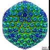
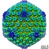
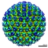
| Title | The HSV-1 UL17 protein is the second constituent of the capsid vertex specific component (CVSC) required for DNA packaging and retention. | |||||||||
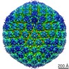
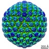
 Map data Map data | Surface rendered view of HSV-1 C-capsid | |||||||||
 Sample Sample |
| |||||||||
 Keywords Keywords | HSV-1 / UL17 / UL25 / CVSC / cryo-EM / virus structure | |||||||||
| Biological species |  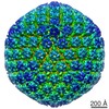 Human herpesvirus 1 strain KOS Human herpesvirus 1 strain KOS | |||||||||
| Method | single particle reconstruction / cryo EM / Resolution: 16.5 Å | |||||||||
 Authors Authors | Toropova K / Huffman JB / Homa FL / Conway JF | |||||||||
 Citation Citation | Journal: J Virol / Year: 2011 Title: The herpes simplex virus 1 UL17 protein is the second constituent of the capsid vertex-specific component required for DNA packaging and retention. Authors: Katerina Toropova / Jamie B Huffman / Fred L Homa / James F Conway /  Abstract: The herpes simplex virus (HSV) UL17 and UL25 minor capsid proteins are essential for DNA packaging. They are thought to comprise a molecule arrayed in five copies around each of the capsid vertices. ...The herpes simplex virus (HSV) UL17 and UL25 minor capsid proteins are essential for DNA packaging. They are thought to comprise a molecule arrayed in five copies around each of the capsid vertices. This molecule was initially termed the "C-capsid-specific component" (CCSC) (B. L. Trus et al., Mol. Cell 26:479-489, 2007), but as we have subsequently observed this feature on reconstructions of A, B, and C capsids, we now refer to it more generally as the "capsid vertex-specific component" (CVSC) (S. K. Cockrell et al., J. Virol. 85:4875-4887, 2011). We previously confirmed that UL25 occupies the vertex-distal region of the CVSC density by visualizing a large UL25-specific tag in reconstructions calculated from cryo-electron microscopy (cryo-EM) images. We have pursued the same strategy to determine the capsid location of the UL17 protein. Recombinant viruses were generated that contained either a small tandem affinity purification (TAP) tag or the green fluorescent protein (GFP) attached to the C terminus of UL17. Purification of the TAP-tagged UL17 or a similarly TAP-tagged UL25 protein clearly demonstrated that the two proteins interact. A cryo-EM reconstruction of capsids containing the UL17-GFP protein reveals that UL17 is the second component of the CVSC and suggests that UL17 interfaces with the other CVSC component, UL25, through its C terminus. The portion of UL17 nearest the vertex appears to be poorly constrained, which may provide flexibility in interacting with tegument proteins or the DNA-packaging machinery at the portal vertex. The exposed locations of the UL17 and UL25 proteins on the HSV-1 capsid exterior suggest that they may be attractive targets for highly specific antivirals. | |||||||||
| History |
|
- Structure visualization
| Movie |
 Movie viewer Movie viewer |
|---|---|
| Structure viewer | EM map: SurfViewMolmilJmol/JSmol |
| Supplemental images |

- Downloads & links
-EMDB archive
| Map data | emd_1902.map.gz | 541.7 MB | EMDB map data format | |
|---|---|---|---|---|
| Header (meta data) | emd-1902-v30.xmlemd-1902.xml | 8.2 KB 8.2 KB | Display Display | EMDB header |
| Images | 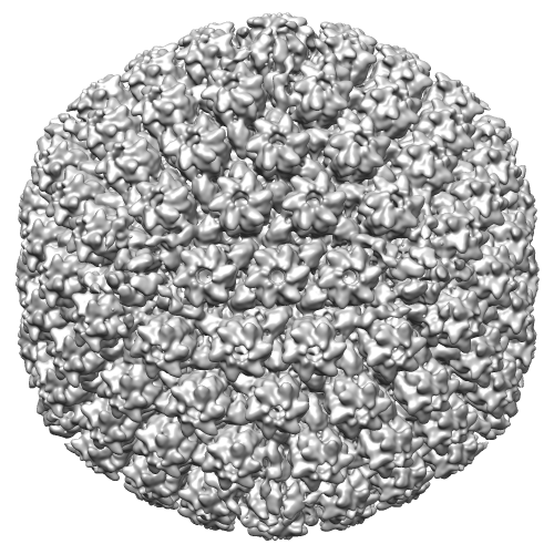 EMD-1902.png EMD-1902.png | 243.6 KB | ||
| Archive directory |  http://ftp.pdbj.org/pub/emdb/structures/EMD-1902ftp://ftp.pdbj.org/pub/emdb/structures/EMD-1902 http://ftp.pdbj.org/pub/emdb/structures/EMD-1902ftp://ftp.pdbj.org/pub/emdb/structures/EMD-1902 | HTTPS FTP |
-Related structure data

-Links
| EMDB pages | EMDB (EBI/PDBe) / EMDataResource |
|---|
-Map
| File | Download / File: emd_1902.map.gz / Format: CCP4 / Size: 985.7 MB / Type: IMAGE STORED AS FLOATING POINT NUMBER (4 BYTES) | ||||||||||||||||||||||||||||||||||||||||||||||||||||||||||||||||||||
|---|---|---|---|---|---|---|---|---|---|---|---|---|---|---|---|---|---|---|---|---|---|---|---|---|---|---|---|---|---|---|---|---|---|---|---|---|---|---|---|---|---|---|---|---|---|---|---|---|---|---|---|---|---|---|---|---|---|---|---|---|---|---|---|---|---|---|---|---|---|
| Annotation | Surface rendered view of HSV-1 C-capsid | ||||||||||||||||||||||||||||||||||||||||||||||||||||||||||||||||||||
| Projections & slices | Image control
Images are generated by Spider. | ||||||||||||||||||||||||||||||||||||||||||||||||||||||||||||||||||||
| Voxel size | X=Y=Z: 2.12 Å | ||||||||||||||||||||||||||||||||||||||||||||||||||||||||||||||||||||
| Density |
| ||||||||||||||||||||||||||||||||||||||||||||||||||||||||||||||||||||
| Symmetry | Space group: 1 | ||||||||||||||||||||||||||||||||||||||||||||||||||||||||||||||||||||
| Details | EMDB XML:
CCP4 map header:
| ||||||||||||||||||||||||||||||||||||||||||||||||||||||||||||||||||||
 Z (Sec.)
 Y (Row.)
 X (Col.)


-Supplemental data
- Sample components
-Entire : HSV-1 wild type C-capsid (KOS).
| Entire | Name: HSV-1 wild type C-capsid (KOS). |
|---|---|
| Components |
|
-Supramolecule #1000: HSV-1 wild type C-capsid (KOS).
| Supramolecule | Name: HSV-1 wild type C-capsid (KOS). / type: sample / ID: 1000 / Number unique components: 1 |
|---|
-Supramolecule #1: Human herpesvirus 1 strain KOS
| Supramolecule | Name: Human herpesvirus 1 strain KOS / type: virus / ID: 1 / Name.synonym: HSV-1 / NCBI-ID: 10306 / Sci species name: Human herpesvirus 1 strain KOS / Virus type: VIRION / Virus isolate: STRAIN / Virus enveloped: No / Virus empty: No / Syn species name: HSV-1 |
|---|---|
| Host (natural) | Organism:  Homo sapiens (human) / synonym: VERTEBRATES Homo sapiens (human) / synonym: VERTEBRATES |
| Virus shell | Shell ID: 1 / Diameter: 1250 Å / T number (triangulation number): 16 |
-Experimental details
-Structure determination
| Method | cryo EM |
|---|---|
 Processing Processing | single particle reconstruction |
| Aggregation state | particle |
-Sample preparation
| Buffer | pH: 7.5 / Details: 500 mM NaCl, 10 mM Tris, 1 mM EDTA |
|---|---|
| Vitrification | Cryogen name: ETHANE / Chamber humidity: 85 % / Instrument: FEI VITROBOT MARK III / Details: Vitrification instrument: Vitrobot mark III / Method: 7.5 second blot before plunging |
- Electron microscopy
| Microscope | FEI TECNAI F20 |
|---|---|
| Date | Jul 7, 2010 |
| Image recording | Category: FILM / Film or detector model: KODAK SO-163 FILM / Digitization - Scanner: NIKON SUPER COOLSCAN 9000 / Digitization - Sampling interval: 6.35 µm / Number real images: 48 / Bits/pixel: 8 |
| Electron beam | Acceleration voltage: 200 kV / Electron source:  FIELD EMISSION GUN FIELD EMISSION GUN |
| Electron optics | Calibrated magnification: 30000 / Illumination mode: FLOOD BEAM / Imaging mode: BRIGHT FIELD / Cs: 2.0 mm / Nominal defocus max: 2.73 µm / Nominal defocus min: 1.63 µm / Nominal magnification: 29000 |
| Sample stage | Specimen holder: Eucentric / Specimen holder model: GATAN LIQUID NITROGEN |
| Experimental equipment |  Model: Tecnai F20 / Image courtesy: FEI Company |
-Image processing
| CTF correction | Details: Phase flip each particle |
|---|---|
| Final reconstruction | Applied symmetry - Point group: I (icosahedral) / Algorithm: OTHER / Resolution.type: BY AUTHOR / Resolution: 16.5 Å / Resolution method: FSC 0.5 CUT-OFF / Software - Name: AUTO3DEM / Number images used: 3973 |
