plus-end-directed organelle transport along microtubule / histone-dependent DNA binding / negative regulation of establishment of protein localization to mitochondrion / neurofibrillary tangle / microtubule lateral binding / axonal transport / tubulin complex / positive regulation of protein localization to synapse / phosphatidylinositol bisphosphate binding / negative regulation of tubulin deacetylation ...plus-end-directed organelle transport along microtubule / histone-dependent DNA binding / negative regulation of establishment of protein localization to mitochondrion / neurofibrillary tangle / microtubule lateral binding / axonal transport / tubulin complex / positive regulation of protein localization to synapse / phosphatidylinositol bisphosphate binding / negative regulation of tubulin deacetylation / generation of neurons / rRNA metabolic process / axonal transport of mitochondrion / regulation of mitochondrial fission / axon development / regulation of chromosome organization / central nervous system neuron development / intracellular distribution of mitochondria / minor groove of adenine-thymine-rich DNA binding / lipoprotein particle binding / microtubule polymerization / negative regulation of mitochondrial membrane potential / regulation of microtubule polymerization / dynactin binding / apolipoprotein binding / main axon / protein polymerization / glial cell projection / axolemma / negative regulation of mitochondrial fission / Caspase-mediated cleavage of cytoskeletal proteins / regulation of microtubule polymerization or depolymerization / neurofibrillary tangle assembly / positive regulation of axon extension / Activation of AMPK downstream of NMDARs / regulation of cellular response to heat / positive regulation of superoxide anion generation / synapse assembly / supramolecular fiber organization / regulation of long-term synaptic depression / positive regulation of protein localization / cellular response to brain-derived neurotrophic factor stimulus / regulation of calcium-mediated signaling / cytoplasmic microtubule organization / positive regulation of microtubule polymerization / somatodendritic compartment / axon cytoplasm / astrocyte activation / stress granule assembly / phosphatidylinositol binding / nuclear periphery / regulation of microtubule cytoskeleton organization / protein phosphatase 2A binding / cellular response to reactive oxygen species / Hsp90 protein binding / microglial cell activation / synapse organization / PKR-mediated signaling / cellular response to nerve growth factor stimulus / protein homooligomerization / regulation of synaptic plasticity / SH3 domain binding / response to lead ion / microtubule cytoskeleton organization / memory / cytoplasmic ribonucleoprotein granule / neuron projection development / cell-cell signaling / single-stranded DNA binding / protein-folding chaperone binding / actin binding / cellular response to heat / growth cone / microtubule cytoskeleton / cell body / double-stranded DNA binding / protein-macromolecule adaptor activity / microtubule binding / dendritic spine / sequence-specific DNA binding / amyloid fibril formation / microtubule / learning or memory / neuron projection / regulation of autophagy / membrane raft / axon / negative regulation of gene expression / neuronal cell body / dendrite / DNA damage response / protein kinase binding / enzyme binding / mitochondrion / DNA binding / RNA binding / extracellular region / identical protein binding / nucleus / plasma membrane 類似検索 - 分子機能
Microtubule-associated protein Tau / Microtubule associated protein, tubulin-binding repeat / Tau and MAP protein, tubulin-binding repeat / Tau and MAP proteins tubulin-binding repeat signature. / Tau and MAP proteins tubulin-binding repeat profile. / : 類似検索 - ドメイン・相同性
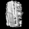
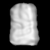
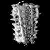
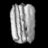
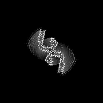
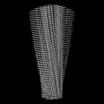
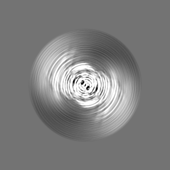
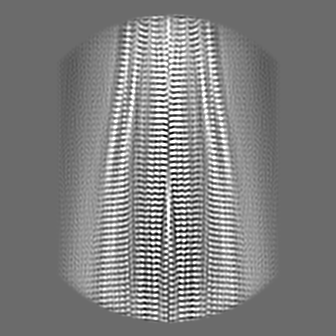
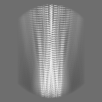
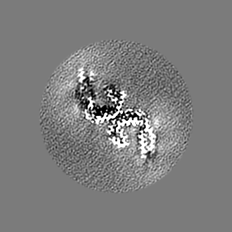
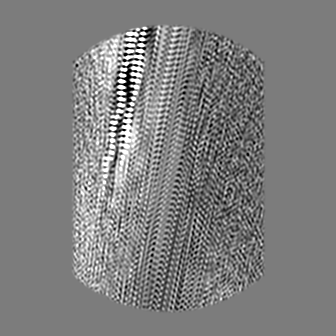
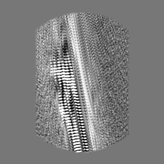
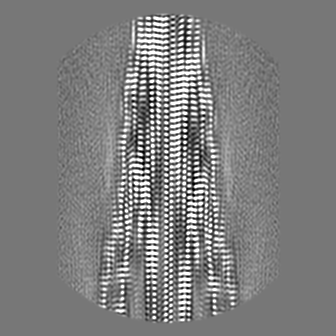
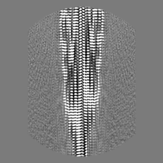
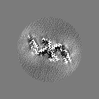
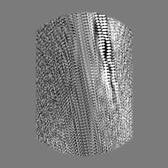
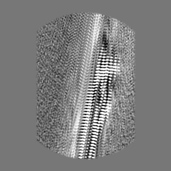
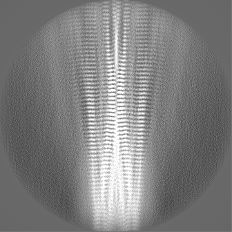
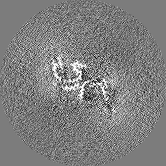
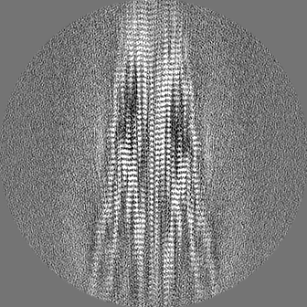
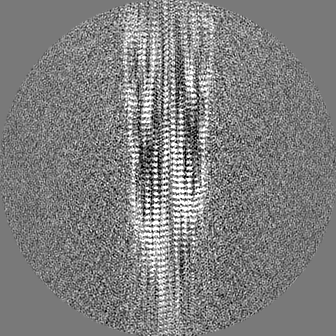
ジャーナル: Nature / 年: 2024 タイトル: CryoET of β-amyloid and tau within postmortem Alzheimer's disease brain. 著者: Madeleine A G Gilbert / Nayab Fatima / Joshua Jenkins / Thomas J O'Sullivan / Andreas Schertel / Yehuda Halfon / Martin Wilkinson / Tjado H J Morrema / Mirjam Geibel / Randy J Read / Neil A ...著者: Madeleine A G Gilbert / Nayab Fatima / Joshua Jenkins / Thomas J O'Sullivan / Andreas Schertel / Yehuda Halfon / Martin Wilkinson / Tjado H J Morrema / Mirjam Geibel / Randy J Read / Neil A Ranson / Sheena E Radford / Jeroen J M Hoozemans / René A W Frank / 要旨: A defining pathological feature of most neurodegenerative diseases is the assembly of proteins into amyloid that form disease-specific structures. In Alzheimer's disease, this is characterized by the ...A defining pathological feature of most neurodegenerative diseases is the assembly of proteins into amyloid that form disease-specific structures. In Alzheimer's disease, this is characterized by the deposition of β-amyloid and tau with disease-specific conformations. The in situ structure of amyloid in the human brain is unknown. Here, using cryo-fluorescence microscopy-targeted cryo-sectioning, cryo-focused ion beam-scanning electron microscopy lift-out and cryo-electron tomography, we determined in-tissue architectures of β-amyloid and tau pathology in a postmortem Alzheimer's disease donor brain. β-amyloid plaques contained a mixture of fibrils, some of which were branched, and protofilaments, arranged in parallel arrays and lattice-like structures. Extracellular vesicles and cuboidal particles defined the non-amyloid constituents of β-amyloid plaques. By contrast, tau inclusions formed parallel clusters of unbranched filaments. Subtomogram averaging a cluster of 136 tau filaments in a single tomogram revealed the polypeptide backbone conformation and filament polarity orientation of paired helical filaments within tissue. Filaments within most clusters were similar to each other, but were different between clusters, showing amyloid heterogeneity that is spatially organized by subcellular location. The in situ structural approaches outlined here for human donor tissues have applications to a broad range of neurodegenerative diseases.
凍結剤: ETHANE / チャンバー内湿度: 90 % / チャンバー内温度: 279 K / 装置: FEI VITROBOT MARK IV
詳細
Frozen brain section was homogenised and purified using sarkosyl detergent.
-
電子顕微鏡法
顕微鏡
FEI TITAN KRIOS
撮影
フィルム・検出器のモデル: FEI FALCON IV (4k x 4k) デジタル化 - サイズ - 横: 4096 pixel / デジタル化 - サイズ - 縦: 4096 pixel / 撮影したグリッド数: 1 / 実像数: 10860 / 平均露光時間: 4.0 sec. / 平均電子線量: 44.0 e/Å2 詳細: Each EER movie was fractionated and tif compressed into 38 frames with a dose of 1.16 e-/A2/frame prior to processing
選択した数: 667095 / ソフトウェア - 名称: crYOLO (ver. 1.8) 詳細: 100 micrographs manually picked and used to train crYOLO model to pick all micrographs, focusing on wider tau filaments rather than thinner abeta
 ムービー
ムービー コントローラー


 データを開く
 基本情報
基本情報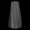
 マップデータ
 試料
試料 キーワード
キーワード 機能・相同性情報
 Homo sapiens (ヒト)
 データ登録者
 英国, 4件
  引用
引用

 構造の表示
構造の表示
 ダウンロードとリンク
 emd_18990.png
 http://ftp.pdbj.org/pub/emdb/structures/EMD-18990


 Z (Sec.)
 Y (Row.)
 X (Col.)


 試料の構成要素
 解析
解析 電子顕微鏡法
 FIELD EMISSION GUN