 Movie
Movie Controller


+ Open data

- Basic information
| Entry | Database: EMDB / ID: EMD-1887 | |||||||||
|---|---|---|---|---|---|---|---|---|---|---|
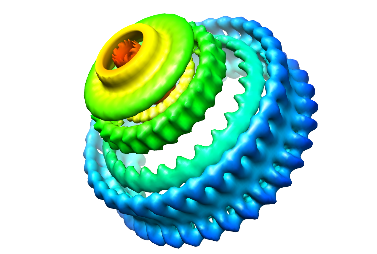
| Title | Flagellar basal body - Salmonella Typhimurium. | |||||||||
 Map data Map data | 34-fold C ring 25-fold M ring for the basal body of S. Typhimurium. | |||||||||
 Sample Sample |
| |||||||||
 Keywords Keywords | bacterial flagellum / Salmonella / C-ring / M-ring / basal body / FliF / FliG | |||||||||
| Biological species |  Salmonella enterica subsp. enterica serovar Typhimurium (bacteria) Salmonella enterica subsp. enterica serovar Typhimurium (bacteria) | |||||||||
| Method | single particle reconstruction / cryo EM / negative staining | |||||||||
 Authors Authors | Thomas DR / Francis NR / Xu C / DeRosier DJ | |||||||||
 Citation Citation | Journal: J Bacteriol / Year: 2006 Title: The three-dimensional structure of the flagellar rotor from a clockwise-locked mutant of Salmonella enterica serovar Typhimurium. Authors: Dennis R Thomas / Noreen R Francis / Chen Xu / David J DeRosier /  Abstract: Three-dimensional reconstructions from electron cryomicrographs of the rotor of the flagellar motor reveal that the symmetry of individual M rings varies from 24-fold to 26-fold while that of the C ...Three-dimensional reconstructions from electron cryomicrographs of the rotor of the flagellar motor reveal that the symmetry of individual M rings varies from 24-fold to 26-fold while that of the C rings, containing the two motor/switch proteins FliM and FliN, varies from 32-fold to 36-fold, with no apparent correlation between the symmetries of the two rings. Results from other studies provided evidence that, in addition to the transmembrane protein FliF, at least some part of the third motor/switch protein, FliG, contributes to a thickening on the face of the M ring, but there was no evidence as to whether or not any portion of FliG also contributes to the C ring. Of the four morphological features in the cross section of the C ring, the feature closest to the M ring is not present with the rotational symmetry of the rest of the C ring, but instead it has the symmetry of the M ring. We suggest that this inner feature arises from a domain of FliG. We present a hypothetical docking in which the C-terminal motor domain of FliG lies in the C ring, where it can interact intimately with FliM. | |||||||||
| History |
|
- Structure visualization
| Movie |
 Movie viewer Movie viewer |
|---|---|
| Structure viewer | EM map: SurfViewMolmilJmol/JSmol |
| Supplemental images |
- Downloads & links
-EMDB archive
| Map data | emd_1887.map.gz | 1.9 MB | EMDB map data format | |
|---|---|---|---|---|
| Header (meta data) | emd-1887-v30.xmlemd-1887.xml | 8.9 KB 8.9 KB | Display Display | EMDB header |
| Images |  1887.png 1887.png | 527.2 KB | ||
| Archive directory |  http://ftp.pdbj.org/pub/emdb/structures/EMD-1887ftp://ftp.pdbj.org/pub/emdb/structures/EMD-1887 http://ftp.pdbj.org/pub/emdb/structures/EMD-1887ftp://ftp.pdbj.org/pub/emdb/structures/EMD-1887 | HTTPS FTP |
-Related structure data
| Similar structure data |
|---|

-Links
| EMDB pages | EMDB (EBI/PDBe) / EMDataResource |
|---|
-Map
| File | Download / File: emd_1887.map.gz / Format: CCP4 / Size: 14.5 MB / Type: IMAGE STORED AS FLOATING POINT NUMBER (4 BYTES) | ||||||||||||||||||||||||||||||||||||||||||||||||||||||||||||||||||||
|---|---|---|---|---|---|---|---|---|---|---|---|---|---|---|---|---|---|---|---|---|---|---|---|---|---|---|---|---|---|---|---|---|---|---|---|---|---|---|---|---|---|---|---|---|---|---|---|---|---|---|---|---|---|---|---|---|---|---|---|---|---|---|---|---|---|---|---|---|---|
| Annotation | 34-fold C ring 25-fold M ring for the basal body of S. Typhimurium. | ||||||||||||||||||||||||||||||||||||||||||||||||||||||||||||||||||||
| Projections & slices | Image control
Images are generated by Spider. generated in cubic-lattice coordinate | ||||||||||||||||||||||||||||||||||||||||||||||||||||||||||||||||||||
| Voxel size | X=Y=Z: 2.8 Å | ||||||||||||||||||||||||||||||||||||||||||||||||||||||||||||||||||||
| Density |
| ||||||||||||||||||||||||||||||||||||||||||||||||||||||||||||||||||||
| Symmetry | Space group: 1 | ||||||||||||||||||||||||||||||||||||||||||||||||||||||||||||||||||||
| Details | EMDB XML:
CCP4 map header:
| ||||||||||||||||||||||||||||||||||||||||||||||||||||||||||||||||||||
 Z (Sec.)
 Y (Row.)
 X (Col.)


-Supplemental data
- Sample components
-Entire : Isolated hook-basal body
| Entire | Name: Isolated hook-basal body |
|---|---|
| Components |
|
-Supramolecule #1000: Isolated hook-basal body
| Supramolecule | Name: Isolated hook-basal body / type: sample / ID: 1000 / Number unique components: 4 |
|---|
-Supramolecule #1: Basal body
| Supramolecule | Name: Basal body / type: organelle_or_cellular_component / ID: 1 / Name.synonym: Rotor / Details: Frozen-hydrated / Oligomeric state: 34-mer C ring, 25-mer M ring / Recombinant expression: Yes |
|---|---|
| Source (natural) | Organism: Salmonella enterica subsp. enterica serovar Typhimurium (bacteria) synonym: Salmonella Typhimurium / Location in cell: Plasma membrane and cytoplasm |
| Recombinant expression | Organism: Salmonella (bacteria) |
-Experimental details
-Structure determination
| Method | negative staining, cryo EM |
|---|---|
 Processing Processing | single particle reconstruction |
| Aggregation state | particle |
-Sample preparation
| Buffer | pH: 8 / Details: 10mM Tris-HCl, 5 mM EDTA, 0.1% Triton X-100 |
|---|---|
| Staining | Type: NEGATIVE Details: Sample put onto Quantifoil grids(R1.2/1.3), blotted with no. 1 Whatman filter paper, plunged into liquid nitrogen-cooled liquid ethane. |
| Grid | Details: Quantifoil grids R1.2/1.3 |
| Vitrification | Cryogen name: ETHANE / Chamber temperature: 100 K / Instrument: HOMEMADE PLUNGER / Details: Vitrification instrument: Home made |
- Electron microscopy
| Microscope | FEI TECNAI F20 |
|---|---|
| Temperature | Average: 100 K |
| Image recording | Category: FILM / Film or detector model: KODAK SO-163 FILM / Digitization - Scanner: ZEISS SCAI / Digitization - Sampling interval: 7 µm / Average electron dose: 10 e/Å2 / Od range: 2 / Bits/pixel: 8 |
| Electron beam | Acceleration voltage: 200 kV / Electron source:  FIELD EMISSION GUN FIELD EMISSION GUN |
| Electron optics | Illumination mode: FLOOD BEAM / Imaging mode: BRIGHT FIELD / Nominal defocus max: 4.0 µm / Nominal magnification: 50000 |
| Sample stage | Specimen holder: Eucentric / Specimen holder model: GATAN LIQUID NITROGEN |
| Experimental equipment |  Model: Tecnai F20 / Image courtesy: FEI Company |
-Image processing
| Details | The images were sorted into classes. 20 classes depending on symmetry of M ring (23 to 26) and symmetry of C ring (32 to 36). |
|---|---|
| CTF correction | Details: CTFFIND, HELIXBOXER |
| Final reconstruction | Software - Name: SPIDER / Number images used: 90 |
