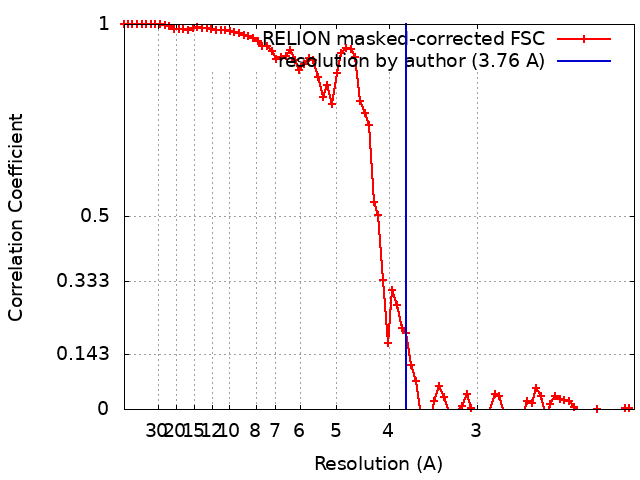
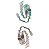
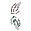
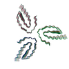
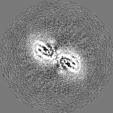
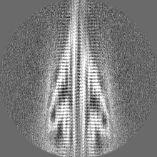
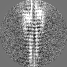
ジャーナル: Nat Commun / 年: 2024 タイトル: Cryo-EM observation of the amyloid key structure of polymorphic TDP-43 amyloid fibrils. 著者: Kartikay Sharma / Fabian Stockert / Jayakrishna Shenoy / Mélanie Berbon / Muhammed Bilal Abdul-Shukkoor / Birgit Habenstein / Antoine Loquet / Matthias Schmidt / Marcus Fändrich / 要旨: The transactive response DNA-binding protein-43 (TDP-43) is a multi-facet protein involved in phase separation, RNA-binding, and alternative splicing. In the context of neurodegenerative diseases, ...The transactive response DNA-binding protein-43 (TDP-43) is a multi-facet protein involved in phase separation, RNA-binding, and alternative splicing. In the context of neurodegenerative diseases, abnormal aggregation of TDP-43 has been linked to amyotrophic lateral sclerosis and frontotemporal lobar degeneration through the aggregation of its C-terminal domain. Here, we report a cryo-electron microscopy (cryo-EM)-based structural characterization of TDP-43 fibrils obtained from the full-length protein. We find that the fibrils are polymorphic and contain three different amyloid structures. The structures differ in the number and relative orientation of the protofilaments, although they share a similar fold containing an amyloid key motif. The observed fibril structures differ from previously described conformations of TDP-43 fibrils and help to better understand the structural landscape of the amyloid fibril structures derived from this protein.
 ムービー
ムービー コントローラー


 データを開く
 基本情報
基本情報
 マップデータ
 試料
試料 キーワード
キーワード 機能・相同性情報
 Homo sapiens (ヒト)
 データ登録者
 引用
引用

 構造の表示
構造の表示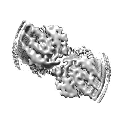
 ダウンロードとリンク
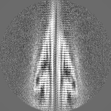
 emd_18715.png
 http://ftp.pdbj.org/pub/emdb/structures/EMD-18715

 Z (Sec.)
 Y (Row.)
 X (Col.)


 試料の構成要素
 解析
解析 電子顕微鏡法
 FIELD EMISSION GUN