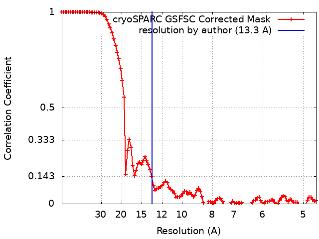
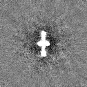
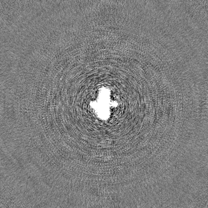
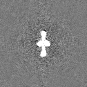
Journal: Int J Mol Sci / Year: 2023 Title: The Structure and Nucleotide-Binding Characteristics of Regulated Cystathionine β-Synthase Domain-Containing Pyrophosphatase without One Catalytic Domain. Authors: Ilya M Zamakhov / Viktor A Anashkin / Andrey V Moiseenko / Victor N Orlov / Natalia N Vorobyeva / Olga S Sokolova / Alexander A Baykov / Abstract: Regulatory adenine nucleotide-binding cystathionine β-synthase (CBS) domains are widespread in proteins; however, information on the mechanism of their modulating effects on protein function is ...Regulatory adenine nucleotide-binding cystathionine β-synthase (CBS) domains are widespread in proteins; however, information on the mechanism of their modulating effects on protein function is scarce. The difficulty in obtaining structural data for such proteins is ascribed to their unusual flexibility and propensity to form higher-order oligomeric structures. In this study, we deleted the most movable domain from the catalytic part of a CBS domain-containing bacterial inorganic pyrophosphatase (CBS-PPase) and characterized the deletion variant both structurally and functionally. The truncated CBS-PPase was inactive but retained the homotetrameric structure of the full-size enzyme and its ability to bind a fluorescent AMP analog (inhibitor) and diadenosine tetraphosphate (activator) with the same or greater affinity. The deletion stabilized the protein structure against thermal unfolding, suggesting that the deleted domain destabilizes the structure in the full-size protein. A "linear" 3D structure with an unusual type of domain swapping predicted for the truncated CBS-PPase by Alphafold2 was confirmed by single-particle electron microscopy. The results suggest a dual role for the CBS domains in CBS-PPase regulation: they allow for enzyme tetramerization, which impedes the motion of one catalytic domain, and bind adenine nucleotides to mitigate or aggravate this effect.
In the structure databanks used in Yorodumi, some data are registered as the other names, "COVID-19 virus" and "2019-nCoV". Here are the details of the virus and the list of structure data.
Jan 31, 2019. EMDB accession codes are about to change! (news from PDBe EMDB page)
EMDB accession codes are about to change! (news from PDBe EMDB page)
The allocation of 4 digits for EMDB accession codes will soon come to an end. Whilst these codes will remain in use, new EMDB accession codes will include an additional digit and will expand incrementally as the available range of codes is exhausted. The current 4-digit format prefixed with “EMD-” (i.e. EMD-XXXX) will advance to a 5-digit format (i.e. EMD-XXXXX), and so on. It is currently estimated that the 4-digit codes will be depleted around Spring 2019, at which point the 5-digit format will come into force.
The EM Navigator/Yorodumi systems omit the EMD- prefix.
Related info.:Q: What is EMD? / ID/Accession-code notation in Yorodumi/EM Navigator
Yorodumi is a browser for structure data from EMDB, PDB, SASBDB, etc.
This page is also the successor to EM Navigator detail page, and also detail information page/front-end page for Omokage search.
The word "yorodu" (or yorozu) is an old Japanese word meaning "ten thousand". "mi" (miru) is to see.
Related info.:EMDB / PDB / SASBDB / Comparison of 3 databanks / Yorodumi Search / Aug 31, 2016. New EM Navigator & Yorodumi / Yorodumi Papers / Jmol/JSmol / Function and homology information / Changes in new EM Navigator and Yorodumi
 Movie
Movie Controller

 Yorodumi
 Open data
 Basic information
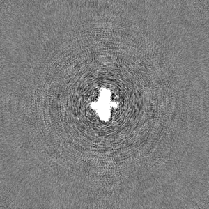
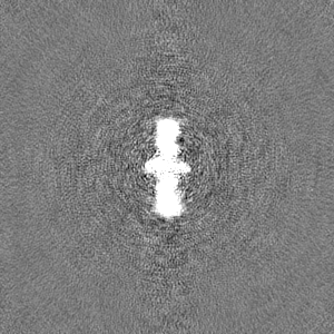
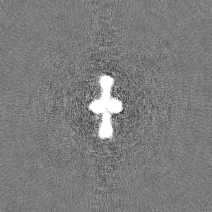
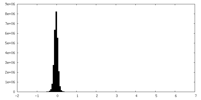
 Map data
 Sample
 Keywords
 Function and homology information
 Desulfitobacterium hafniense DCB-2 (bacteria)
 Authors
 Russian Federation, 1 items
  Citation
 Structure visualization
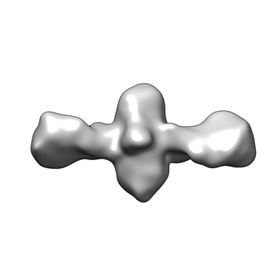
 Downloads & links
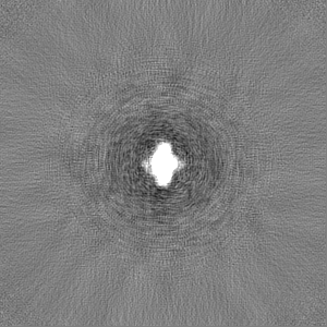
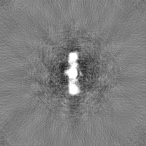
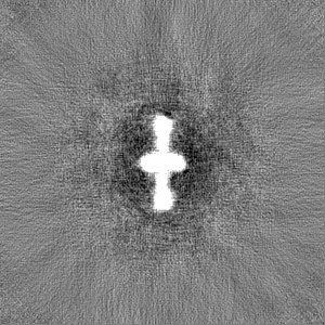
 emd_18687.png
 http://ftp.pdbj.org/pub/emdb/structures/EMD-18687
 Z (Sec.)
 Y (Row.)
 X (Col.)


 Sample components
 Processing
 Electron microscopy