Movie
Movie Controller

[English] 日本語
 Yorodumi
- EMDB-18586: Cryo-EM reconstruction of crosslinked native Bacillus subtilis co... -
+ Open data

- Basic information
| Entry |  | |||||||||
|---|---|---|---|---|---|---|---|---|---|---|
| Title | Cryo-EM reconstruction of crosslinked native Bacillus subtilis collided disome binding MutS2 SMR and KOW domains | |||||||||
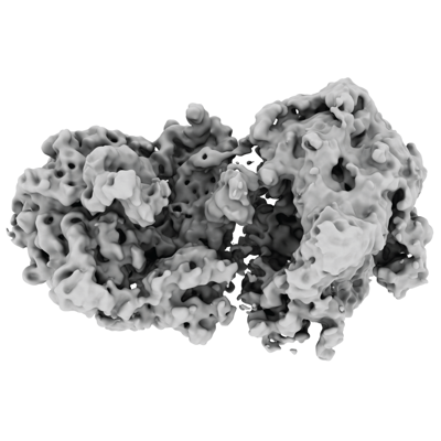
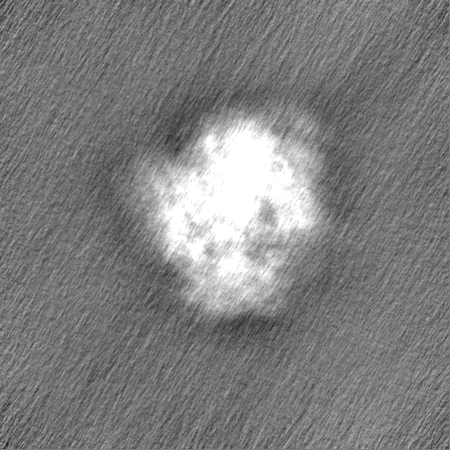
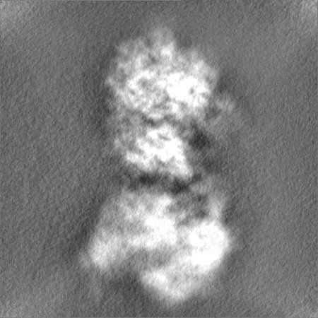
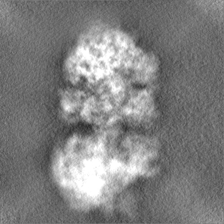
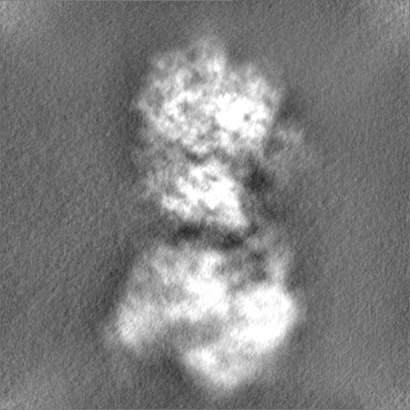
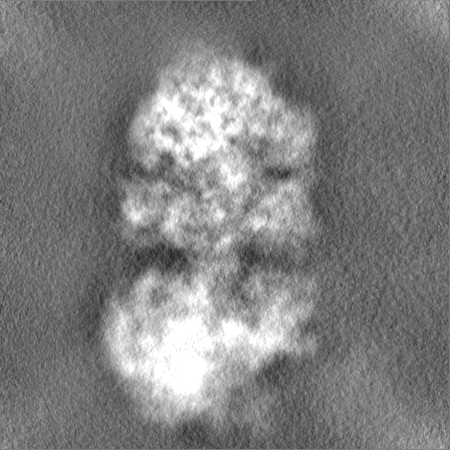
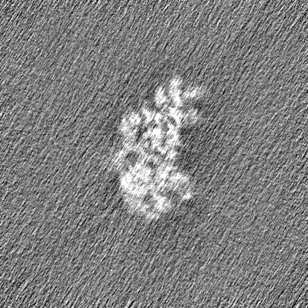
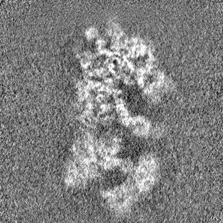
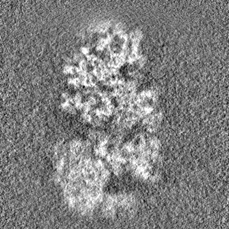
 Map data Map data | Cryo-EM reconstruction of B. subtilis MutS2-disome complex | |||||||||
 Sample Sample |
| |||||||||
 Keywords Keywords | Muts2 / collision / disome / splitting / quality control / RIBOSOME | |||||||||
| Biological species |  | |||||||||
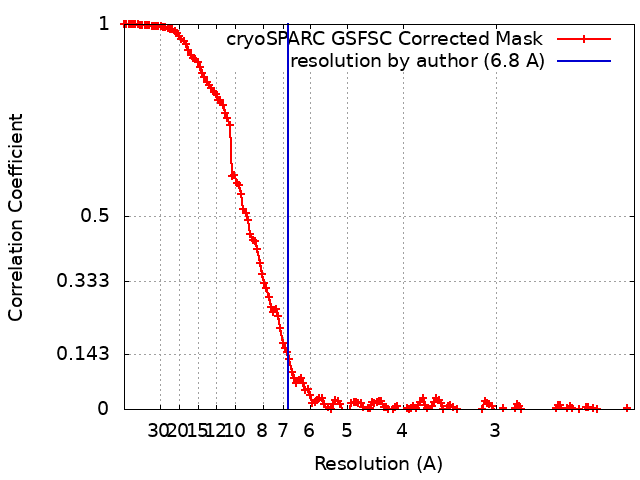
| Method | single particle reconstruction / cryo EM / Resolution: 6.8 Å | |||||||||
 Authors Authors | Park E / Mackens-Kiani T / Berhane R / Esser H / Erdenebat C / Burroughs AM / Berninghausen O / Aravind L / Beckmann R / Green R / Buskirk AR | |||||||||
| Funding support |  Germany, 1 items Germany, 1 items
| |||||||||
 Citation Citation | Journal: EMBO J / Year: 2024 Title: B. subtilis MutS2 splits stalled ribosomes into subunits without mRNA cleavage. Authors: Esther N Park / Timur Mackens-Kiani / Rebekah Berhane / Hanna Esser / Chimeg Erdenebat / A Maxwell Burroughs / Otto Berninghausen / L Aravind / Roland Beckmann / Rachel Green / Allen R Buskirk /  Abstract: Stalled ribosomes are rescued by pathways that recycle the ribosome and target the nascent polypeptide for degradation. In E. coli, these pathways are triggered by ribosome collisions through the ...Stalled ribosomes are rescued by pathways that recycle the ribosome and target the nascent polypeptide for degradation. In E. coli, these pathways are triggered by ribosome collisions through the recruitment of SmrB, a nuclease that cleaves the mRNA. In B. subtilis, the related protein MutS2 was recently implicated in ribosome rescue. Here we show that MutS2 is recruited to collisions by its SMR and KOW domains, and we reveal the interaction of these domains with collided ribosomes by cryo-EM. Using a combination of in vivo and in vitro approaches, we show that MutS2 uses its ABC ATPase activity to split ribosomes, targeting the nascent peptide for degradation through the ribosome quality control pathway. However, unlike SmrB, which cleaves mRNA in E. coli, we see no evidence that MutS2 mediates mRNA cleavage or promotes ribosome rescue by tmRNA. These findings clarify the biochemical and cellular roles of MutS2 in ribosome rescue in B. subtilis and raise questions about how these pathways function differently in diverse bacteria. | |||||||||
| History |
|
- Structure visualization
| Supplemental images |
|---|
- Downloads & links
-EMDB archive
| Map data | emd_18586.map.gz | 171.6 MB |  EMDB map data format EMDB map data format | |
|---|---|---|---|---|
| Header (meta data) | emd-18586-v30.xmlemd-18586.xml | 19.7 KB 19.7 KB | Display Display | EMDB header |
| FSC (resolution estimation) | emd_18586_fsc.xml | 15.1 KB | Display | FSC data file |
| Images |  emd_18586.png emd_18586.png | 122.5 KB | ||
| Filedesc metadata | emd-18586.cif.gz | 4 KB | ||
| Others | emd_18586_half_map_1.map.gzemd_18586_half_map_2.map.gz | 322.3 MB 322.3 MB | ||
| Archive directory |  http://ftp.pdbj.org/pub/emdb/structures/EMD-18586ftp://ftp.pdbj.org/pub/emdb/structures/EMD-18586 http://ftp.pdbj.org/pub/emdb/structures/EMD-18586ftp://ftp.pdbj.org/pub/emdb/structures/EMD-18586 | HTTPS FTP |
-Validation report
| Summary document | emd_18586_validation.pdf.gz | 1.2 MB | Display | EMDB validaton report |
|---|---|---|---|---|
| Full document | emd_18586_full_validation.pdf.gz | 1.2 MB | Display | |
| Data in XML | emd_18586_validation.xml.gz | 24.1 KB | Display | |
| Data in CIF | emd_18586_validation.cif.gz | 31.5 KB | Display | |
| Arichive directory | https://ftp.pdbj.org/pub/emdb/validation_reports/EMD-18586ftp://ftp.pdbj.org/pub/emdb/validation_reports/EMD-18586 | HTTPS FTP |
-Related structure data


-Links
| EMDB pages | EMDB (EBI/PDBe) / EMDataResource |
|---|
-Map
| File | Download / File: emd_18586.map.gz / Format: CCP4 / Size: 347.6 MB / Type: IMAGE STORED AS FLOATING POINT NUMBER (4 BYTES) | ||||||||||||||||||||||||||||||||||||
|---|---|---|---|---|---|---|---|---|---|---|---|---|---|---|---|---|---|---|---|---|---|---|---|---|---|---|---|---|---|---|---|---|---|---|---|---|---|
| Annotation | Cryo-EM reconstruction of B. subtilis MutS2-disome complex | ||||||||||||||||||||||||||||||||||||
| Projections & slices | Image control
Images are generated by Spider. | ||||||||||||||||||||||||||||||||||||
| Voxel size | X=Y=Z: 1.084 Å | ||||||||||||||||||||||||||||||||||||
| Density |
| ||||||||||||||||||||||||||||||||||||
| Symmetry | Space group: 1 | ||||||||||||||||||||||||||||||||||||
| Details | EMDB XML:
|
 Z (Sec.)
 Y (Row.)
 X (Col.)


-Supplemental data
-Half map: Cryo-EM reconstruction of B. subtilis MutS2-disome complex
| File | emd_18586_half_map_1.map | ||||||||||||
|---|---|---|---|---|---|---|---|---|---|---|---|---|---|
| Annotation | Cryo-EM reconstruction of B. subtilis MutS2-disome complex | ||||||||||||
| Projections & Slices |
| ||||||||||||
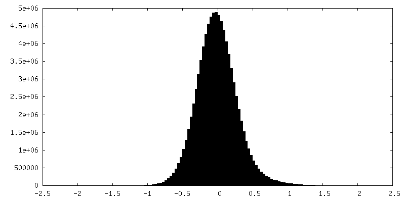
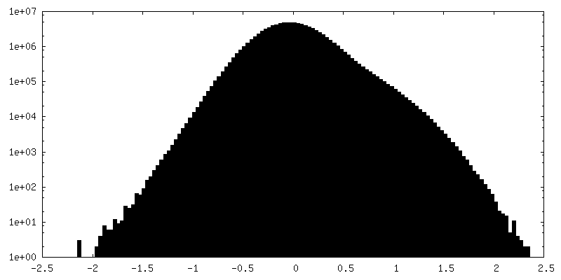
| Density Histograms |


-Half map: Cryo-EM reconstruction of B. subtilis MutS2-disome complex
| File | emd_18586_half_map_2.map | ||||||||||||
|---|---|---|---|---|---|---|---|---|---|---|---|---|---|
| Annotation | Cryo-EM reconstruction of B. subtilis MutS2-disome complex | ||||||||||||
| Projections & Slices |
| ||||||||||||
| Density Histograms |
- Sample components
-Entire : MutS2-disome complex from B. subtilis, focus refined, stalled 70S
| Entire | Name: MutS2-disome complex from B. subtilis, focus refined, stalled 70S |
|---|---|
| Components |
|
-Supramolecule #1: MutS2-disome complex from B. subtilis, focus refined, stalled 70S
| Supramolecule | Name: MutS2-disome complex from B. subtilis, focus refined, stalled 70S type: complex / ID: 1 / Parent: 0 / Macromolecule list: #1-#6, #51, #53, #7-#50, #52 |
|---|---|
| Source (natural) | Organism: |
-Experimental details
-Structure determination
| Method | cryo EM |
|---|---|
 Processing Processing | single particle reconstruction |
| Aggregation state | particle |
-Sample preparation
| Buffer | pH: 7.5 |
|---|---|
| Vitrification | Cryogen name: ETHANE |
- Electron microscopy
| Microscope | FEI TITAN KRIOS |
|---|---|
| Image recording | Film or detector model: GATAN K2 SUMMIT (4k x 4k) / Average electron dose: 43.6 e/Å2 |
| Electron beam | Acceleration voltage: 300 kV / Electron source:  FIELD EMISSION GUN FIELD EMISSION GUN |
| Electron optics | Illumination mode: FLOOD BEAM / Imaging mode: BRIGHT FIELD / Nominal defocus max: 3.5 µm / Nominal defocus min: 0.4 µm |
| Experimental equipment |  Model: Titan Krios / Image courtesy: FEI Company |