 Movie
Movie Controller

[English] 日本語
 Yorodumi
- EMDB-1856: Automatic Recovery of Missing Amplitudes and Phases in Tilt-Limit... -
+ Open data

- Basic information
| Entry | Database: EMDB / ID: EMD-1856 | |||||||||
|---|---|---|---|---|---|---|---|---|---|---|
| Title | Automatic Recovery of Missing Amplitudes and Phases in Tilt-Limited Electron Crystallography of 2D Crystals | |||||||||
 Map data Map data | Crystal unit cell of 3D-PCO reconstructed bacteriorhodopsin. Generated from original MTZ limited from 15.0 A to 2.5 A. | |||||||||
 Sample Sample |
| |||||||||
 Keywords Keywords | Crystallography / Signal Recovery / Iterative Methods / Oversampling / Tilt-Limited / Electron Microscopy | |||||||||
| Biological species |  Halobacterium salinarum (Halophile) Halobacterium salinarum (Halophile) | |||||||||
| Method | electron crystallography / cryo EM | |||||||||
 Authors Authors | Gipson B / Masiel DJ / Browning ND / Spence J / Mitsuoka K / Stahlberg H | |||||||||
 Citation Citation | Journal: Phys Rev E Stat Nonlin Soft Matter Phys / Year: 2011 Title: Automatic recovery of missing amplitudes and phases in tilt-limited electron crystallography of two-dimensional crystals. Authors: Bryant R Gipson / Daniel J Masiel / Nigel D Browning / John Spence / Kaoru Mitsuoka / Henning Stahlberg /  Abstract: Electron crystallography of 2D protein crystals provides a powerful tool for the determination of membrane protein structure. In this method, data is acquired in the Fourier domain as randomly ...Electron crystallography of 2D protein crystals provides a powerful tool for the determination of membrane protein structure. In this method, data is acquired in the Fourier domain as randomly sampled, uncoupled, amplitudes and phases. Due to physical constraints on specimen tilting, those Fourier data show a vast un-sampled "missing cone" of information, producing resolution loss in the direction perpendicular to the membrane plane. Based on the flexible language of projection onto sets, we provide a full solution for these problems with a projective constraint optimization algorithm that, for sufficiently oversampled data, produces complete recovery of unmeasured data in the missing cone. We apply this method to an experimental data set of Bacteriorhodopsin and show that, in addition to producing superior results compared to traditional reconstruction methods, full, reproducible, recovery of the missing cone from noisy data is possible. Finally, we present an automatic implementation of the refinement routine as open source, freely distributed, software that will be included in our 2dx software package. | |||||||||
| History |
|
- Structure visualization
| Movie |
 Movie viewer Movie viewer |
|---|---|
| Structure viewer | EM map: SurfViewMolmilJmol/JSmol |
| Supplemental images |
 UCSF Chimera
- Downloads & links
-EMDB archive
| Map data | emd_1856.map.gz | 3.5 MB | EMDB map data format | |
|---|---|---|---|---|
| Header (meta data) | emd-1856-v30.xmlemd-1856.xml | 7.5 KB 7.5 KB | Display Display | EMDB header |
| Images | 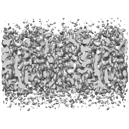 EMD-1856.png EMD-1856.png | 193.6 KB | ||
| Archive directory |  http://ftp.pdbj.org/pub/emdb/structures/EMD-1856ftp://ftp.pdbj.org/pub/emdb/structures/EMD-1856 http://ftp.pdbj.org/pub/emdb/structures/EMD-1856ftp://ftp.pdbj.org/pub/emdb/structures/EMD-1856 | HTTPS FTP |
-Links
| EMDB pages | EMDB (EBI/PDBe) / EMDataResource |
|---|---|
| Related items in Molecule of the Month |

-Map
| File | Download / File: emd_1856.map.gz / Format: CCP4 / Size: 3.7 MB / Type: IMAGE STORED AS FLOATING POINT NUMBER (4 BYTES) | ||||||||||||||||||||||||||||||||||||||||||||||||||||||||||||||||||||
|---|---|---|---|---|---|---|---|---|---|---|---|---|---|---|---|---|---|---|---|---|---|---|---|---|---|---|---|---|---|---|---|---|---|---|---|---|---|---|---|---|---|---|---|---|---|---|---|---|---|---|---|---|---|---|---|---|---|---|---|---|---|---|---|---|---|---|---|---|---|
| Annotation | Crystal unit cell of 3D-PCO reconstructed bacteriorhodopsin. Generated from original MTZ limited from 15.0 A to 2.5 A. | ||||||||||||||||||||||||||||||||||||||||||||||||||||||||||||||||||||
| Projections & slices | Image control
Images are generated by Spider. | ||||||||||||||||||||||||||||||||||||||||||||||||||||||||||||||||||||
| Voxel size | X: 0.82171 Å / Y: 0.82171 Å / Z: 1 Å | ||||||||||||||||||||||||||||||||||||||||||||||||||||||||||||||||||||
| Density |
| ||||||||||||||||||||||||||||||||||||||||||||||||||||||||||||||||||||
| Symmetry | Space group: 1 | ||||||||||||||||||||||||||||||||||||||||||||||||||||||||||||||||||||
| Details | EMDB XML:
CCP4 map header:
| ||||||||||||||||||||||||||||||||||||||||||||||||||||||||||||||||||||
 Z (Sec.)
 Y (Row.)
 X (Col.)


-Supplemental data
- Sample components
-Entire : Bacteriorhodopsin
| Entire | Name: Bacteriorhodopsin |
|---|---|
| Components |
|
-Supramolecule #1000: Bacteriorhodopsin
| Supramolecule | Name: Bacteriorhodopsin / type: sample / ID: 1000 / Details: Exactly as per PDB entry 1at9 (same dataset) / Number unique components: 1 |
|---|
-Macromolecule #1: Bacteriorhodopsin
| Macromolecule | Name: Bacteriorhodopsin / type: protein_or_peptide / ID: 1 / Name.synonym: BR / Recombinant expression: No / Database: NCBI |
|---|---|
| Source (natural) | Organism: Halobacterium salinarum (Halophile) |
-Experimental details
-Structure determination
| Method | cryo EM |
|---|---|
 Processing Processing | electron crystallography |
| Aggregation state | 2D array |
-Sample preparation
| Vitrification | Cryogen name: NITROGEN / Chamber temperature: 4.2 K / Instrument: OTHER |
|---|
- Electron microscopy
| Microscope | JEOL KYOTO-3000SFF |
|---|---|
| Details | The full details of the original dataset can be found from PDB entry 1at9 |
| Electron beam | Acceleration voltage: 300 kV / Electron source: OTHER |
| Electron optics | Illumination mode: OTHER / Imaging mode: OTHER |
| Sample stage | Specimen holder: As per 1at9 / Specimen holder model: JEOL |
-Image processing
| Final reconstruction | Algorithm: OTHER / Software - Name: 2dx (PCO Module) Details: 3D-PCO reconstructed maps, generated from phase-flipped raw 2D kimura, 97 crystallographic dataset. |
|---|---|
| Crystal parameters | Plane group: P 3 |
| CTF correction | Details: Phase flipping and Electron Diffraction |
