 Movie
Movie Controller


+ Open data

- Basic information
| Entry | Database: EMDB / ID: EMD-1846 | |||||||||
|---|---|---|---|---|---|---|---|---|---|---|
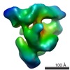
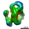
| Title | Human native spliceosomal C complex assembled on PM5 pre-mRNA. | |||||||||
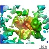
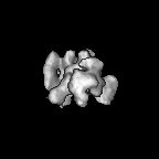
 Map data Map data | This is a 3D map of the human spliceosomal C complex. | |||||||||
 Sample Sample |
| |||||||||
 Keywords Keywords | spliceosome / pre-mRNA splicing / C complex / catalytically active / PM5 pre-mRNA | |||||||||
| Biological species |  Homo sapiens (human) Homo sapiens (human) | |||||||||
| Method | single particle reconstruction / cryo EM / Resolution: 29.0 Å | |||||||||
 Authors Authors | Golas MM / Sander B / Bessonov S / Grote M / Wolf E / Kastner B / Stark H / Luhrmann R | |||||||||
 Citation Citation | Journal: Mol Cell / Year: 2010 Title: 3D cryo-EM structure of an active step I spliceosome and localization of its catalytic core. Authors: Monika M Golas / Bjoern Sander / Sergey Bessonov / Michael Grote / Elmar Wolf / Berthold Kastner / Holger Stark / Reinhard Lührmann /  Abstract: The spliceosome excises introns from pre-mRNA in a two-step splicing reaction. So far, the three-dimensional (3D) structure of a spliceosome with preserved catalytic activity has remained elusive. ...The spliceosome excises introns from pre-mRNA in a two-step splicing reaction. So far, the three-dimensional (3D) structure of a spliceosome with preserved catalytic activity has remained elusive. Here, we determined the 3D structure of the human, catalytically active step I spliceosome (C complex) by cryo-electron microscopy (cryo-EM) in vitrified ice. Via immunolabeling we mapped the position of the 5' exon. The C complex contains an unusually salt-stable ribonucleoprotein (RNP) core that harbors its catalytic center. We determined the 3D structure of this RNP core and also that of a post-step II particle, the 35S U5 snRNP, which contains most of the C complex core proteins. As C complex domains could be recognized in these structures, their position in the C complex could be determined, thereby allowing the region harboring the spliceosome's catalytic core to be localized. | |||||||||
| History |
|
- Structure visualization
| Movie |
 Movie viewer Movie viewer |
|---|---|
| Structure viewer | EM map: SurfViewMolmilJmol/JSmol |
| Supplemental images |

- Downloads & links
-EMDB archive
| Map data | emd_1846.map.gz | 3.8 MB | EMDB map data format | |
|---|---|---|---|---|
| Header (meta data) | emd-1846-v30.xmlemd-1846.xml | 6.6 KB 6.6 KB | Display Display | EMDB header |
| Images |  emd_1846.png emd_1846.png | 89.6 KB | ||
| Archive directory |  http://ftp.pdbj.org/pub/emdb/structures/EMD-1846ftp://ftp.pdbj.org/pub/emdb/structures/EMD-1846 http://ftp.pdbj.org/pub/emdb/structures/EMD-1846ftp://ftp.pdbj.org/pub/emdb/structures/EMD-1846 | HTTPS FTP |
-Related structure data


-Links
| EMDB pages | EMDB (EBI/PDBe) / EMDataResource |
|---|
-Map
| File | Download / File: emd_1846.map.gz / Format: CCP4 / Size: 11.1 MB / Type: IMAGE STORED AS FLOATING POINT NUMBER (4 BYTES) | ||||||||||||||||||||||||||||||||||||||||||||||||||||||||||||||||||||
|---|---|---|---|---|---|---|---|---|---|---|---|---|---|---|---|---|---|---|---|---|---|---|---|---|---|---|---|---|---|---|---|---|---|---|---|---|---|---|---|---|---|---|---|---|---|---|---|---|---|---|---|---|---|---|---|---|---|---|---|---|---|---|---|---|---|---|---|---|---|
| Annotation | This is a 3D map of the human spliceosomal C complex. | ||||||||||||||||||||||||||||||||||||||||||||||||||||||||||||||||||||
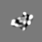
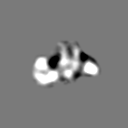
| Projections & slices | Image control
Images are generated by Spider. | ||||||||||||||||||||||||||||||||||||||||||||||||||||||||||||||||||||
| Voxel size | X=Y=Z: 4.9 Å | ||||||||||||||||||||||||||||||||||||||||||||||||||||||||||||||||||||
| Density |
| ||||||||||||||||||||||||||||||||||||||||||||||||||||||||||||||||||||
| Symmetry | Space group: 1 | ||||||||||||||||||||||||||||||||||||||||||||||||||||||||||||||||||||
| Details | EMDB XML:
CCP4 map header:
| ||||||||||||||||||||||||||||||||||||||||||||||||||||||||||||||||||||
 Z (Sec.)
 Y (Row.)
 X (Col.)


-Supplemental data
- Sample components
-Entire : Human native spliceosomal C complex
| Entire | Name: Human native spliceosomal C complex |
|---|---|
| Components |
|
-Supramolecule #1000: Human native spliceosomal C complex
| Supramolecule | Name: Human native spliceosomal C complex / type: sample / ID: 1000 / Details: Experimental weight in the range of 5-5.5MDa / Number unique components: 1 |
|---|---|
| Molecular weight | Experimental: 5 MDa |
-Supramolecule #1: Human native spliceosomal C complex
| Supramolecule | Name: Human native spliceosomal C complex / type: organelle_or_cellular_component / ID: 1 / Name.synonym: Spliceosomal C complex / Recombinant expression: No |
|---|---|
| Source (natural) | Organism: Homo sapiens (human) / synonym: Human |
-Experimental details
-Structure determination
| Method | cryo EM |
|---|---|
 Processing Processing | single particle reconstruction |
| Aggregation state | particle |
-Sample preparation
| Vitrification | Cryogen name: ETHANE / Instrument: OTHER |
|---|
- Electron microscopy
| Microscope | FEI/PHILIPS CM200FEG |
|---|---|
| Electron beam | Acceleration voltage: 160 kV / Electron source:  FIELD EMISSION GUN FIELD EMISSION GUN |
| Electron optics | Illumination mode: SPOT SCAN / Imaging mode: BRIGHT FIELD |
| Sample stage | Specimen holder: Eucentric / Specimen holder model: GATAN LIQUID NITROGEN |
-Image processing
| Final reconstruction | Applied symmetry - Point group: C1 (asymmetric) / Resolution.type: BY AUTHOR / Resolution: 29.0 Å / Resolution method: FSC 0.5 CUT-OFF |
|---|
