 Movie
Movie Controller


+ Open data

- Basic information
| Entry |  | ||||||||||||||||||||||||||||||||||||
|---|---|---|---|---|---|---|---|---|---|---|---|---|---|---|---|---|---|---|---|---|---|---|---|---|---|---|---|---|---|---|---|---|---|---|---|---|---|
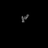
| Title | cryo-EM map of apo Clostridioides difficile toxin B | ||||||||||||||||||||||||||||||||||||
 Map data Map data | |||||||||||||||||||||||||||||||||||||
 Sample Sample |
| ||||||||||||||||||||||||||||||||||||
 Keywords Keywords | microbiology / pore forming region / CROP dynamics / TOXIN | ||||||||||||||||||||||||||||||||||||
| Biological species |  Clostridioides difficile (bacteria) Clostridioides difficile (bacteria) | ||||||||||||||||||||||||||||||||||||
| Method | single particle reconstruction / cryo EM / Resolution: 3.21 Å | ||||||||||||||||||||||||||||||||||||
 Authors Authors | Kinsolving J / Bous J | ||||||||||||||||||||||||||||||||||||
| Funding support |  Sweden, Sweden,  Denmark, 11 items Denmark, 11 items
| ||||||||||||||||||||||||||||||||||||
 Citation Citation | Journal: Cell Rep / Year: 2024 Title: Structural and functional insight into the interaction of Clostridioides difficile toxin B and FZD. Authors: Julia Kinsolving / Julien Bous / Pawel Kozielewicz / Sara Košenina / Rawan Shekhani / Lukas Grätz / Geoffrey Masuyer / Yuankai Wang / Pål Stenmark / Min Dong / Gunnar Schulte /  Abstract: The G protein-coupled receptors of the Frizzled (FZD) family, in particular FZD, are receptors that are exploited by Clostridioides difficile toxin B (TcdB), the major virulence factor responsible ...The G protein-coupled receptors of the Frizzled (FZD) family, in particular FZD, are receptors that are exploited by Clostridioides difficile toxin B (TcdB), the major virulence factor responsible for pathogenesis associated with Clostridioides difficile infection. We employ a live-cell assay examining the affinity between full-length FZDs and TcdB. Moreover, we present cryoelectron microscopy structures of TcdB alone and in complex with full-length FZD, which reveal that large structural rearrangements of the combined repetitive polypeptide domain are required for interaction with FZDs and other TcdB receptors, constituting a first step for receptor recognition. Furthermore, we show that bezlotoxumab, an FDA-approved monoclonal antibody to treat Clostridioides difficile infection, favors the apo-TcdB structure and thus disrupts binding with FZD. The dynamic transition between the two conformations of TcdB also governs the stability of the pore-forming region. Thus, our work provides structural and functional insight into how conformational dynamics of TcdB determine receptor binding. | ||||||||||||||||||||||||||||||||||||
| History |
|
- Structure visualization
| Supplemental images |
|---|

- Downloads & links
-EMDB archive
| Map data | emd_18403.map.gz | 240.6 MB |  EMDB map data format EMDB map data format | |
|---|---|---|---|---|
| Header (meta data) | emd-18403-v30.xmlemd-18403.xml | 22 KB 22 KB | Display Display | EMDB header |
| FSC (resolution estimation) | emd_18403_fsc.xmlemd_18403_fsc_2.xml | 16.5 KB 16.5 KB | Display Display | FSC data file |
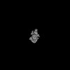
| Images |  emd_18403.png emd_18403.png | 77.1 KB | ||
| Filedesc metadata | emd-18403.cif.gz | 7 KB | ||
| Others | emd_18403_half_map_1.map.gzemd_18403_half_map_2.map.gz | 442.1 MB 442.1 MB | ||
| Archive directory |  http://ftp.pdbj.org/pub/emdb/structures/EMD-18403ftp://ftp.pdbj.org/pub/emdb/structures/EMD-18403 http://ftp.pdbj.org/pub/emdb/structures/EMD-18403ftp://ftp.pdbj.org/pub/emdb/structures/EMD-18403 | HTTPS FTP |
-Related structure data


-Links
| EMDB pages | EMDB (EBI/PDBe) / EMDataResource |
|---|
-Map
| File | Download / File: emd_18403.map.gz / Format: CCP4 / Size: 476.8 MB / Type: IMAGE STORED AS FLOATING POINT NUMBER (4 BYTES) | ||||||||||||||||||||||||||||||||||||
|---|---|---|---|---|---|---|---|---|---|---|---|---|---|---|---|---|---|---|---|---|---|---|---|---|---|---|---|---|---|---|---|---|---|---|---|---|---|
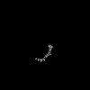
| Projections & slices | Image control
Images are generated by Spider. | ||||||||||||||||||||||||||||||||||||
| Voxel size | X=Y=Z: 1.1592 Å | ||||||||||||||||||||||||||||||||||||
| Density |
| ||||||||||||||||||||||||||||||||||||
| Symmetry | Space group: 1 | ||||||||||||||||||||||||||||||||||||
| Details | EMDB XML:
|
 Z (Sec.)
 Y (Row.)
 X (Col.)


-Supplemental data
-Half map: #2
| File | emd_18403_half_map_1.map | ||||||||||||
|---|---|---|---|---|---|---|---|---|---|---|---|---|---|
| Projections & Slices |
| ||||||||||||
| Density Histograms |


-Half map: #1
| File | emd_18403_half_map_2.map | ||||||||||||
|---|---|---|---|---|---|---|---|---|---|---|---|---|---|
| Projections & Slices |
| ||||||||||||
| Density Histograms |


- Sample components
-Entire : apo-structure of Clostridioides difficile toxin B
| Entire | Name: apo-structure of Clostridioides difficile toxin B |
|---|---|
| Components |
|
-Supramolecule #1: apo-structure of Clostridioides difficile toxin B
| Supramolecule | Name: apo-structure of Clostridioides difficile toxin B / type: organelle_or_cellular_component / ID: 1 / Parent: 0 / Macromolecule list: all |
|---|---|
| Source (natural) | Organism: Clostridioides difficile (bacteria) |
-Supramolecule #2: masked region-GTD/CPD domain
| Supramolecule | Name: masked region-GTD/CPD domain / type: organelle_or_cellular_component / ID: 2 / Parent: 1 / Macromolecule list: all |
|---|---|
| Source (natural) | Organism: Clostridioides difficile (bacteria) |
-Macromolecule #1: apo-TcdB
| Macromolecule | Name: apo-TcdB / type: other / ID: 1 / Classification: other |
|---|---|
| Source (natural) | Organism: Clostridioides difficile (bacteria) |
| Sequence | String: mdklvhlnqr gkctmslvnr kqlekmanvr frtqedeyva ildaleeyhn msentvveky lklkdinslt diyidtykks grnkalkkfk eylvtevl e lknnnltpve knlhfvwigg qindtainyi nqwkdvnsdy nvnvfydsna flintlkktv vesaindtle ...String: mdklvhlnqr gkctmslvnr kqlekmanvr frtqedeyva ildaleeyhn msentvveky lklkdinslt diyidtykks grnkalkkfk eylvtevl e lknnnltpve knlhfvwigg qindtainyi nqwkdvnsdy nvnvfydsna flintlkktv vesaindtle sfrenlndpr fdynkffrkr meiiydk qk nfinyykaqr eenpeliidd ivktylsney skeidelnty ieeslnkitq nsgndvrnfe efkngesfnl yeqelverwn laaasdilri salkeigg m yldvdmlpgi qpdlfesiek pssvtvdfwe mtkleaimky keyipeytse hfdmldeevq ssfesvlask sdkseifssl gdmeasplev kiaf nskgi inqglisvkd sycsnlivkq ienrykilnn slnpaisedn dfntttntfi dsimaeanad ngrfmmelgk ylrvgffpdv kttinlsgpe ayaaa yqdl lmfkegsmni hlieadlrnf eisktnisqs teqemaslws fddarakaqf eeykrnyfeg slgeddnldf sqnivvdkey llekisslar sserg yihy ivqlqgdkis yeaacnlfak tpydsvlfqk niedseiayy ynpgdgeiqe idkykipsii sdrpkikltf ighgkdefnt difagfdvds lsteieaa i dlakedispk sieinllgcn mfsysinvee typgklllkv kdkiselmps isqdsiivsa nqyevrinse grrelldhsg ewinkeesii kdisskeyis f npkenkit vksknlpels tllqeirnns nssdieleek vmlteceinv isnidtqive erieeaknlt sdsinyikde fkliesisda lcdlkqqnel edsh fisfe disetdegfs irfinketge sifvetekti fseyanhite eiskikgtif dtvngklvkk vnldtthevn tlnaaffiqs lieynsskes lsnlsvamkv qvyaqlfst glntitdaak vvelvstald etidllptls eglpiiatii dgvslgaaik elsetsdpll rqeieakigi mavnlttatt aiitsslgia sgfsillvpl a gisagips lvnnelvlrd katkvvdyfk hvslvetegv ftllddkimm pqddlvisei dfnnnsivlg kceiwrmegg sghtvtddid hffsapsity r ephlsiyd vlevqkeeld lskdlmvlpn apnrvfawet gwtpglrsle ndgtklldri rdnyegefyw ryfafiadal ittlkpryed tnirinldsn trs fivpii tteyirekls ysfygsggty alslsqynmg inielsesdv wiidvdnvvr dvtiesdkik kgdliegils tlsieenkii lnsheinfsg evngsngfv sltfsilegi naiievdlls ksykllisge lkilmlnsnh iqqkidyigf nselqknipy sfvdsegken gfingstkeg lfvselpdvv liskvymdds kpsf gyysn nlkdvkvitk dnvniltgyy lkddikisls ltlqdektik lnsvhldesg vaeilkfmnr kgntntsdsl msflesmnik sifvnflqsn ikfildan f iisgttsigq feficdendn iqpyfikfnt letnytlyvg nrqnmivepn ydlddsgdis stvinfsqky lygidscvnk vvispniytd einitpvyet n ntypeviv ldanyineki nvnindlsir yvwsndgndf ilmstseenk vsqvkirfvn vfkdktlank lsfnfsdkqd vpvseiilsf tpsyyedgli gy dlglvsl ynekfyinnf gmmvsgliyi ndslyyfkpp vnnlitgfvt vgddkyyfnp inggaasige tiiddknyyf nqsgvlqtgv fstedgfkyf apa ntlden legeaidftg kliideniyy fddnyrgave wkeldgemhy fspetgkafk glnqigdyky yfnsdgvmqk gfvsindnkh yfddsgvmk vgyteidgkh fyfaengemq igvfntedgf kyfahhnedl gneegeeisy sgilnfnnki yyfddsftav vgwkdledgs kyyfdedtae ayiglsli n dgqyyfnddg imqvgfvtin dkvfyfsdsg iiesgvqnid dnyfyiddng ivqigvfdts dgykyfapan tvndniygqa veysglvrvg edvyyf get ytietgwiyd menesdkyyf npetkkackg inliddikyy fdekgimrtg lisfennnyy fnengemqfg yiniedkmfy fgedgvmqig vf ntpdgfk yfahqntlde nfegesinyt gwldldekry yftdeyiaat gsviidgeey yfdpdtaqlv isegyrphag lrgshhhhhh |
| Recombinant expression | Organism: Priestia megaterium DSM 319 (bacteria) |
-Experimental details
-Structure determination
| Method | cryo EM |
|---|---|
 Processing Processing | single particle reconstruction |
| Aggregation state | particle |
-Sample preparation
| Concentration | 0.630 mg/mL | ||||||||||||||||||
|---|---|---|---|---|---|---|---|---|---|---|---|---|---|---|---|---|---|---|---|
| Buffer | pH: 7.5 Component:
Details: 100 mM TRIS-HCl pH 7.5 200 mM NaCl 0.002% LMNG 0.0002% CHS 0.0002% GDN | ||||||||||||||||||
| Vitrification | Cryogen name: ETHANE / Chamber humidity: 100 % / Chamber temperature: 277 K / Instrument: FEI VITROBOT MARK IV |
- Electron microscopy
| Microscope | FEI TITAN KRIOS |
|---|---|
| Specialist optics | Energy filter - Slit width: 20 eV |
| Image recording | Film or detector model: GATAN K3 (6k x 4k) / Number grids imaged: 1 / Number real images: 17269 / Average exposure time: 3.0 sec. / Average electron dose: 50.453 e/Å2 Details: Images collected in super-resolution mode, faster acquisition mode, with 4 exposures per hole |
| Electron beam | Acceleration voltage: 300 kV / Electron source:  FIELD EMISSION GUN FIELD EMISSION GUN |
| Electron optics | Calibrated magnification: 105000 / Illumination mode: FLOOD BEAM / Imaging mode: BRIGHT FIELD / Cs: 2.7 mm / Nominal defocus max: 2.0 µm / Nominal defocus min: 0.6 µm |
| Sample stage | Cooling holder cryogen: NITROGEN |
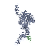
| Experimental equipment |  Model: Titan Krios / Image courtesy: FEI Company |
+Image processing

-Atomic model buiding 1
| Initial model | Chain - Source name: AlphaFold / Chain - Initial model type: in silico model |
|---|---|
| Refinement | Space: REAL / Protocol: RIGID BODY FIT |
